Comparative Study
. 2013 Dec:117:62-78.
doi: 10.1016/j.exer.2013.07.027. Epub 2013 Aug 14.

Tear film mucins: front line defenders of the ocular surface; comparison with airway and gastrointestinal tract mucins

Affiliations
Comparative Study

Tear film mucins: front line defenders of the ocular surface; comparison with airway and gastrointestinal tract mucins

Robin R Hodges et al. Exp Eye Res. 2013 Dec.

Abstract

The ocular surface including the cornea and conjunctiva and its overlying tear film are the first tissues of the eye to interact with the external environment. The tear film is complex containing multiple layers secreted by different glands and tissues. Each layer contains specific molecules and proteins that not only maintain the health of the cells on the ocular surface by providing nourishment and removal of waste products but also protect these cells from environment. A major protective mechanism that the corneal and conjunctival cells have developed is secretion of the innermost layer of the tear film, the mucous layer. Both the cornea and conjunctiva express membrane spanning mucins, whereas the conjunctiva also produces soluble mucins. The mucins present in the tear film serve to maintain the hydration of the ocular surface and to provide lubrication and anti-adhesive properties between the cells of the ocular surface and conjunctiva during the blink. A third function is to contribute to the epithelial barrier to prevent pathogens from binding to the ocular surface. This review will focus on the different types of mucins produced by the corneal and conjunctival epithelia. Also included in this review will be a presentation of the structure of mucins, regulation of mucin production, role of mucins in ocular surface diseases, and the differences in mucin production by the ocular surface, airways and gastrointestinal tract.

Keywords: conjunctiva; cornea; mucin; proliferation; secretion; tears.


Figures

Figure 1
Figure 1
Mucin Producing Tissues of the Ocular Surface. Acinar cells of the lacrimal gland, epithelial cells of the cornea and conjunctiva, and goblet cells of the conjunctiva synthesize and secrete mucins onto the ocular surface. Photographs are electron micrographs of indicated tissue. Micrograph of cornea is reproduced from Gipson and Joyce. Anatomy and Cell Biology of the Cornea, Superficial Limbus; and Conjunctiva in Principles and Practice of Ophthalmology. 2000 Albert and Jakobiec, ed. WB Saunders Company, Philadelphia.
Figure 2
Figure 2
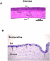
Structure of cornea and conjunctiva. Hematoxylin and eosin staining of human cornea (A). Arrow at the top of tissue indicates epithelium while arrow at bottom of tissue indicates endothelium. Arrowheads indicate keratocytes present in the stroma. Reprinted from Leal and Pearlman, The role of cytokines and pathogen recognition molecules in fungal keratitis – Insights from human disease and animal models. Cytokines 2012 58:107-111. Alcian blue/periodic acid Schiff reagent staining of rat conjunctiva (B). Arrows indicate clusters of several goblet cells. Reprinted from Shatos et al. Isolation, characterization, and propagation of rat conjunctival goblet cells in vitro. Invest Ophthalmol Vis Sci 2001, 42:1455-1464. Epi: epithelium; Endo: endothelium.
Figure 3
Figure 3
Schematic diagram of structures of membrane spanning mucins. CT: cytoplasmic tails; SEA: Sea urchin sperm protein Enterokinase and Agrin module; GPDH: glycerol-3-phosphate dehydrogenase; S: serine residues; T: threonine residues; Y: tyrosine residues. Reprinted from Govindarjan and Gipson. Membrane-tethered mucins have multiple functions on the ocular surface. Exp Eye Res. 2010, 90:655-663.
Figure 4
Figure 4
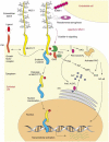
Outsid-in Signaling by MUC1. MUC1 signaling can be mediated by phosphorylation of hte MUC1CT by ligand binding and activation of a growth factor receptor, interactions with ICAM-1 or bacteria binding to activate several signaling pathways. The MUC1CT can be cleaved and translocate to the nucleus and potentially regulate genes. ICAM-1, intracellular adhesion molecule 1; PLCγ, phospholipase Cγ; DAG, diacylglycerol; PKC, protein kinase C; PM, plasma membrane; ER, endoplasmic reticulum; Grb2: growth factor receptor-bound protein; Sos: a guanine nucleotide exchange factor ; Ras: small GTPase; Raf: mitogen-activated protein kinase kinase kinase; MEK: mitogen-activated protein kinase kinase; ERK, extracellular regulated kinase; NF-κB- nuclear factor κB; From Singh and Hollingsworth. Cell surface-associated mucins in signal transduction. Trends in Cell Biol 2006, 16:467-476.
Figure 5
Figure 5
Schematic diagram of the structure of the protein core of the small secretory mucin MUC7. Modified from Gipson and Argueso. Role of Mucins in the Function of the Corneal and Conjunctival Epithelia. Int Rev Cytol. 2003, 231:1-49.
Figure 6
Figure 6
Schematic diagram of the structure of the protein core of the gel-forming mucin MUC5AC. Modified from Gipson and Argueso. Role of Mucins in the Function of the Corneal and Conjunctival Epithelia. Int Rev Cytol. 2003, 231:1-49.
Figure 7
Figure 7
Schematic diagram of EGF signaling pathway to stimulate proliferation of conjunctival goblet cells. PIP2: phosphatidylinositol 4,5-bisphosphate; DAG: diacylglycerol; PKCα: protein kinase Cα; IP3: inositol trisphosphate; ER: endoplasmic reticulum; PLCγ: phospholipase Cγ; PI3K: phosphoinositide 3-kinase; AKT: protein kinase B; EGF: epidermal growth factor; EGFR: epidermal growth factor receptor; Shc: adaptor protein; Grb2: growth factor receptor-bound protein; Sos: a guanine nucleotide exchange factor ; Ras: small GTPase; Raf: mitogen-activated protein kinase kinase kinase; MEK: mitogen-activated protein kinase kinase; MAPK: mitogen-activated protein kinase (also known as extracellular regulated kinase (ERK) 1/2); MEKK-1: mitogen-activated protein kinase kinase kinase 1; MKK7: mitogen-activated protein kinase kinase 7; JNK: c-Jun N-terminal kinase; c-jun: transcription factor; ELK-1: transcription activator.
Figure 8
Figure 8
Schematic diagram of signaling pathways used to stimulate mucin secretion in conjunctival goblet cells. Signaling pathway used by cholinergic agonists (A) or vasoactive intestinal peptide (VIP, B) to stimulate mucin secretion. EGF: epidermal growth factor; EGFR: epidermal growth factor receptor; Ras: small GTPase; Raf: mitogen-activated protein kinase kinase kinase; MEK: mitogen-activated protein kinase kinase; ERK 1/2: extracellular regulated kinase (ERK) 1/2 (also known as mitogen-activated protein kinase (MAPK)); PLCγ: phospholipase Cγ; MMP: matrix metalloproteinase; Pyk2: a nonreceptor tyrosine kinase; Src: a nonreceptor tyrosine kinase; DAG: diacylglycerol; IP3: inositol trisphosphate; M1, M2, M3: muscarinic receptors of the M1, M2, and M3 subtypes; Cch: carbachol; α subunit of Gq/11 G protein; Gαs: α subunit of Gs G protein; VPAC1 and 2: vasoactive intestinal peptide receptors 1 and 2; AC: adenylate cyclase; cAMP: cyclic adenosine monophosphate; PKA: protein kinase A. Reprinted from Li et al Effect of VIP on intracellular [Ca2+], extracellular regulated kinase 1/2, and secretion in cultured rat conjunctival goblet cells. Invest Ophthalmol Vis Sci 2013 23:2872-2884.
Figure 8
Figure 8
Schematic diagram of signaling pathways used to stimulate mucin secretion in conjunctival goblet cells. Signaling pathway used by cholinergic agonists (A) or vasoactive intestinal peptide (VIP, B) to stimulate mucin secretion. EGF: epidermal growth factor; EGFR: epidermal growth factor receptor; Ras: small GTPase; Raf: mitogen-activated protein kinase kinase kinase; MEK: mitogen-activated protein kinase kinase; ERK 1/2: extracellular regulated kinase (ERK) 1/2 (also known as mitogen-activated protein kinase (MAPK)); PLCγ: phospholipase Cγ; MMP: matrix metalloproteinase; Pyk2: a nonreceptor tyrosine kinase; Src: a nonreceptor tyrosine kinase; DAG: diacylglycerol; IP3: inositol trisphosphate; M1, M2, M3: muscarinic receptors of the M1, M2, and M3 subtypes; Cch: carbachol; α subunit of Gq/11 G protein; Gαs: α subunit of Gs G protein; VPAC1 and 2: vasoactive intestinal peptide receptors 1 and 2; AC: adenylate cyclase; cAMP: cyclic adenosine monophosphate; PKA: protein kinase A. Reprinted from Li et al Effect of VIP on intracellular [Ca2+], extracellular regulated kinase 1/2, and secretion in cultured rat conjunctival goblet cells. Invest Ophthalmol Vis Sci 2013 23:2872-2884.
Figure 9
Figure 9
Counter regulation of the H1 histamine receptor by resolvin D1 in conjunctival goblet cells to prevent histamine stimulation of secretion. H1: histamine receptor subtype 1; PLC: phospholipase C; IP3: inositol trisphosphate; ERK 1/2: extracellular regulated kinase (ERK) 1/2 (also known as mitogen-activated protein kinase (MAPK)); pERK1/2; phosphorylated (active) ERK; β-ARK: β-adrenergic receptor kinase (also known as G protein-coupled receptor kinase (GRK) 2); RvD1: resolvin D1; GPR32: G protein-coupled receptor 32); PKC: protein kinase C.

References

    1. Albertsmeyer AC, Kakkassery V, Spurr-Michaud S, Beeks O, Gipson IK. Effect of pro-inflammatory mediators on membrane-associated mucins expressed by human ocular surface epithelial cells. Exp Eye Res. 2010;90:444–451. - PMC - PubMed
    1. Aragona P, Romeo GF, Puzzolo D, Micali A, Ferreri G. Impression cytology of the conjunctival epithelium in patients with vernal conjunctivitis. Eye (Lond) 1996;10(Pt 1):82–85. - PubMed
    1. Argueso P. Glycobiology of the ocular surface: mucins and lectins. Jpn J Ophthalmol. 2013;57:150–155. - PMC - PubMed
    1. Argueso P, Balaram M, Spurr-Michaud S, Keutmann HT, Dana MR, Gipson IK. Decreased levels of the goblet cell mucin MUC5AC in tears of patients with Sjogren syndrome. Invest Ophthalmol Vis Sci. 2002;43:1004–1011. - PubMed
    1. Argueso P, Guzman-Aranguez A, Mantelli F, Cao Z, Ricciuto J, Panjwani N. Association of cell surface mucins with galectin-3 contributes to the ocular surface epithelial barrier. The Journal of biological chemistry. 2009;284:23037–23045. - PMC - PubMed

Publication types