Review
. 2013 Dec 1;531(2):133-49.
doi: 10.1016/j.gene.2013.08.004. Epub 2013 Aug 14.

Kinesin-5: cross-bridging mechanism to targeted clinical therapy

Affiliations
Review

Kinesin-5: cross-bridging mechanism to targeted clinical therapy

Edward J Wojcik et al. Gene. .

Abstract

Kinesin motor proteins comprise an ATPase superfamily that works hand in hand with microtubules in every eukaryote. The mitotic kinesins, by virtue of their potential therapeutic role in cancerous cells, have been a major focus of research for the past 28 years since the discovery of the canonical Kinesin-1 heavy chain. Perhaps the simplest player in mitotic spindle assembly, Kinesin-5 (also known as Kif11, Eg5, or kinesin spindle protein, KSP) is a plus-end-directed motor localized to interpolar spindle microtubules and to the spindle poles. Comprised of a homotetramer complex, its function primarily is to slide anti-parallel microtubules apart from one another. Based on multi-faceted analyses of this motor from numerous laboratories over the years, we have learned a great deal about the function of this motor at the atomic level for catalysis and as an integrated element of the cytoskeleton. These data have, in turn, informed the function of motile kinesins on the whole, as well as spearheaded integrative models of the mitotic apparatus in particular and regulation of the microtubule cytoskeleton in general. We review what is known about how this nanomotor works, its place inside the cytoskeleton of cells, and its small-molecule inhibitors that provide a toolbox for understanding motor function and for anticancer treatment in the clinic.

Keywords: ADP; AMPPNP; AP1; ATP; ATP hydrolysis; Allosteric inhibition; CDK1; DMSO; DNA; Eg5; GFP; GTP; ICL; KHC; KSP; Kinesin; L5; MT; Mitosis; NCBI; NER; NTP; NTPases; National Center for Biotechnology Information; P loop; P(i); PDB; Protein DataBank; RNAi; RefSeq; Reference Sequence; S-trityl-l-cysteine; STC; Traf4; XPF; Xeroderma pigmentosum group F; activator protein 1; adenosine diphosphate; adenosine-5′-(β,γ-imido)triphosphate; adenosine-5′-triphosphate; cryo-EM; cryo-electron microscopy; cyclin-dependent kinase 1; deoxyribonucleic acid; dimethyl sulfoxide; double stranded ribonucleic acid interference; dsRNAi; green fluorescent protein; guanosine-5′-triphosphate; inorganic phosphate; interstrand DNA cross-linking; kDa; kilodalton; kinesin heavy chain; kinesin spindle protein; loop 5; microtubule; nucleotide excision repair; nucleotide triphosphatases; nucleotide triphosphate; pN; phosphate-binding loop; piconewton; ribonucleic acid interference; siRNA; small interfering ribonucleic acid; tumor necrosis factor receptor associated factor 4.


Figures

Figure 1
Figure 1. Phylogenetic relationship between Kinesin-5 proteins
The phylogenetic analysis is shown as a polar dendrogram with individual sequences labels arranged radially. Seventy-four Kinesin-5 motor domain protein sequences were analyzed by the maximum likelihood, co-estimation method, SATé (Liu et al., 2009). The sequences are labeled with an NCBI GI identifier, protein name (if known), residues corresponding to the motor domain, followed by genus and species. Sequences included were identified from kinesin phylogenies (Wickstead et al., 2010) and by the National Center for Biotechnology Information (NCBI) protein database Reference Sequence (RefSeq). The multiple sequence alignment and maximum likelihood phylogeny were co-calculated by SATé (Liu et al., 2009; Liu et al., 2012). SATé called user defined sequence alignment [MAFFT 6.717; (Katoh et al., 2002)], merger [OPAL 1.0.3; (Wheeler and Kececioglu, 2007)], and phylogeny algorithms [FASTTREE 2.1.4; (Price et al., 2010)]. The decomposition strategy was set to centroid with a maximum subproblem size of 37 sequences. Calculations were allowed to run for a total of 20 iterations without improvement in the maximum likelihood score. Following the final iteration, a final RAxML (Stamatakis, 2006) phylogeny was calculated. Final maximum likelihood score for the phylogeny was −22525.88. Fig Tree v1.3 was used to for visualization.
Figure 2
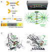
Figure 2. Sequence, function, and structure of Kinesin-5 proteins
(A) Linear sequence organization and general structure of the domains within one kinesin molecule are shown at the bottom of the panel. The asterisk and pound signs highlight the position of the cover neck and the necklinker, respectively. Dimer and tetrameric organization of the Kinesin-5 proteins is also drawn. (B) Cartoon representation of the mitotic spindle and the tetrameric Eg5 molecules cross-bridging spindle microtubules. (C) Two views of the HsEg5 motor domain crystal structure (PDB 3HQD) rotated approximately 120° relative to each other. A non-hydrolyzable AMPPNP molecule bound in the active site. Highlighted are the L5 loop (green), central beta sheet (dark grey), neck linker (black), and the α4 helix (blue). The left and right panels orient the active-site and the microtubule-binding site of HsEg5 to the reader, respectively.
Figure 3
Figure 3. Inhibition of Kinesin-5 by small-molecule inhibitor or knockdown in eukaryotic cells
(A) Chemical structure of monastrol and S-trityl-L-cysteine (STC), which are two allosteric inhibitors of human Kinesin-5. After seeding at a density of 1x10⁶ cells and a 24 hr incubation, human HeLa cells were treated with either (B) DMSO or (C) 1 mM STC. Rounded cell shape is diagnostic of live cells in metaphase. From cell counts, 4% of DMSO-treated cells were in metaphase, whereas 43% of STC-treated cells were in metaphase after a 12 hour-long incubation with this drug. Images (B–C) were acquired using a Nikon ELWD 0.3 phase contrast microscope under 10X magnification. Kinesin-5 (Klp61F) dsRNAi knockdown in Drosophila S2 cells expressing tubulin-GFP prevented morphogenesis of bipolar spindles and, instead, exhibited mono-polar arrays. (D) Confocal fluorescence images of living S2 cells expressing tubulin-GFP after dsRNAi knockdown of native Klp61F. The green (GFP) channel of cells displaying aberrant mono-polar mitosis is shown. (E) Red channel of cells in Panel D showing no detectable expression of Klp61F-mKATE chimera. (F) Merge of panels D and E. (G) Tubulin-GFP expression in Klp61F dsRNAi cells transfected with Klp61F-mKATE chimera. Shown is a confocal image of a rescued bipolar spindle in a living transfected cell. (H) Red channel of cells in panel G showing Klp61F-mKATE localization in transfected cells, with untransfected cells nearby. (I) Merge of panels G and H. Images (D–I) were acquired using a Zeiss Axiovert 200 inverted microscope equipped with a Yokogawa spinning disk confocal accessory. 10 X 63x/1.4 oil DIC.
Figure 4
Figure 4. Experimentally identified phosphorylation sites on Kinesin-5
(A) Phosphosites of HsEg5 (black text) and BMK-1 (grey text) that are required to regulate Kinesin-5 function. (B) Additional phosphosites identified in human, Drosophila, and Xenopus in which their role in regulation is undetermined. Cartoon representation of Kinesin-5 linear sequence (grey rectangle) with phosphorylation sites (green spheres) in approximate location with motor, stalk or tail domains.
Figure 5
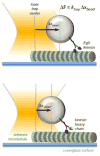
Figure 5. Measuring kinesin stall force with an optical trap
Using a focused laser line, a sole polystyrene bead can be held in place with high precision. With a single kinesin attached by its tail to the bead, the bead can be placed in close enough proximity to a tethered microtubule that the kinesin can bind to, and begin stepping along, the microtubule. As the kinesin pulls the bead away from the trap center, the tendency of the bead to remain in the center exerts a rearward force on the kinesin molecule, which becomes greater as the kinesin attempts to pull the bead further from the center. Thus, force (F) can be measured as a function of distance (x) if the spring constant of the trap (k) is known. Here, we show Eg5 (Kinesin-5) generating a greater force than kinesin heavy chain (Kinesin-1) as evidenced by its ability to pull the bead further from the trap center (Δx), given the same trap stiffness.

Similar articles

Cited by

References

    1. Asenjo AB, Krohn N, Sosa H. Configuration of the two kinesin motor domains during ATP hydrolysis. Nat Struct Biol. 2003;10:836–42. - PubMed
    1. Atherton-Fessler S, Parker LL, Geahlen RL, Piwnica-Worms H. Mechanisms of p34cdc2 regulation. Mol Cell Biol. 1993;13:1675–85. - PMC - PubMed
    1. Avunie-Masala R, Movshovich N, Nissenkorn Y, Gerson-Gurwitz A, Fridman V, Koivomagi M, Loog M, Hoyt MA, Zaritsky A, Gheber L. Phospho-regulation of kinesin-5 during anaphase spindle elongation. J Cell Sci. 2011;124:873–8. - PMC - PubMed
    1. Bannigan A, Scheible WR, Lukowitz W, Fagerstrom C, Wadsworth P, Somerville C, Baskin TI. A conserved role for kinesin-5 in plant mitosis. J Cell Sci. 2007;120:2819–27. - PubMed
    1. Beer T, Goldman B, Synold T, Ryan C, Vasist L, Van Veldhuizen P, Dakhil S, Lara P, Drelichman A, Hussain M, Crawford E. Southwest Oncology Group phase II study of ispinesib in androgen-independent prostate cancer previously treated with taxanes. Clinical Genitourinary Cancer. 2008;6:103–109. - PubMed

Publication types