. 2013 Sep 12;51(5):584-93.
doi: 10.1016/j.molcel.2013.07.025. Epub 2013 Aug 15.

Haemolysin coregulated protein is an exported receptor and chaperone of type VI secretion substrates

Affiliations

Haemolysin coregulated protein is an exported receptor and chaperone of type VI secretion substrates

Julie M Silverman et al. Mol Cell. .

Abstract

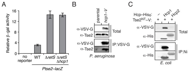
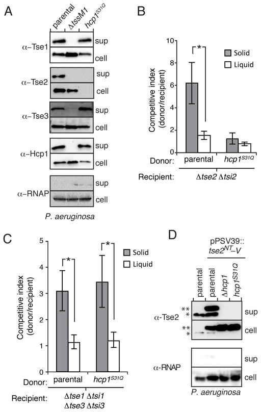
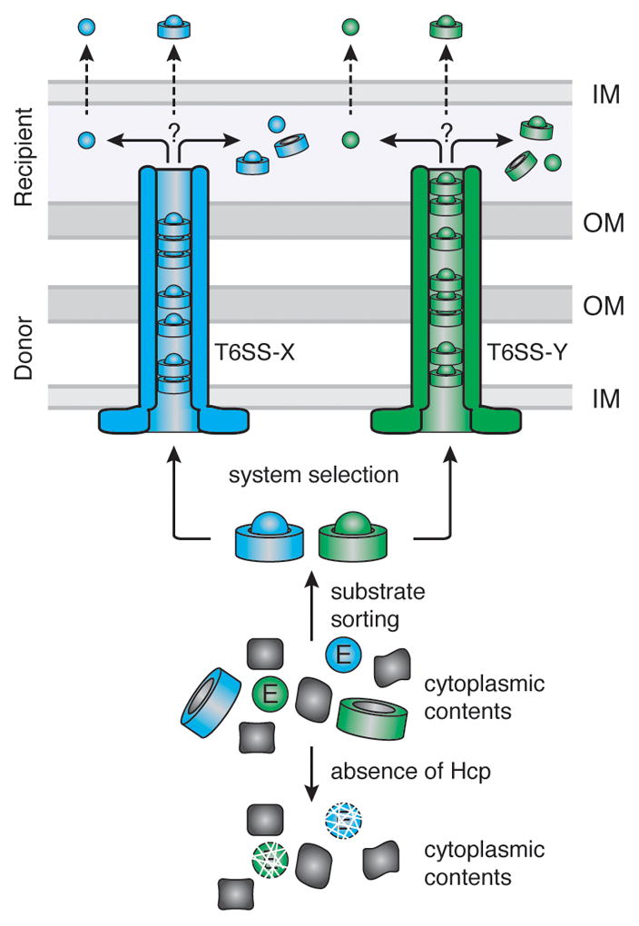
Secretion systems require high-fidelity mechanisms to discriminate substrates among the vast cytoplasmic pool of proteins. Factors mediating substrate recognition by the type VI secretion system (T6SS) of Gram-negative bacteria, a widespread pathway that translocates effector proteins into target bacterial cells, have not been defined. We report that haemolysin coregulated protein (Hcp), a ring-shaped hexamer secreted by all characterized T6SSs, binds specifically to cognate effector molecules. Electron microscopy analysis of an Hcp-effector complex from Pseudomonas aeruginosa revealed the effector bound to the inner surface of Hcp. Further studies demonstrated that interaction with the Hcp pore is a general requirement for secretion of diverse effectors encompassing several enzymatic classes. Though previous models depict Hcp as a static conduit, our data indicate it is a chaperone and receptor of substrates. These unique functions of a secreted protein highlight fundamental differences between the export mechanism of T6 and other characterized secretory pathways.


Figures

Figure 1
Figure 1. Tse2 Requires Hcp1 for Intracellular Accumulation
(A and B) Western blot analysis of intracellular levels of H1-T6S effectors (Tse1, Tse2 and Tse3) in the indicated P. aeruginosa backgrounds. RNA polymerase (RNAP) is included as a loading control. Unless otherwise indicated, the parental background in this and all subsequent figures is ΔretS. (C) Western blot analysis of intracellular Hcp1 and H1-T6S effector levels in a ClpXP-dependent Hcp1 depletion assay. Samples were processed 90 minutes after induced sspB expression in P. aeruginosa strains lacking the native sspB gene and containing wild-type hcp1 or hcp1–D4.
Figure 2
Figure 2. Hcp1 Interacts Directly with Tse2
(A) Relative levels of β-galactosidase activity from the indicated P. aeruginosa strains containing a chromosomally-integrated lacZ reporter fused to the promoter region and first eight codons of tse2 (Ptse2–lacZ). Error bars represent standard deviation based on three independent replicates. (B) Western blot analysis of Tse2 from total and bead-associated fractions of an anti-VSV-G immunoprecipitation from P. aeruginosa strains encoding Hcp1 or ectopically expressing Hcp1. (C) Immunoblot detecting total and bead-associated fractions of a nickel-NTA precipitation assay from E. coli expressing a nontoxic, VSV-G epitope-tagged allele of tse2 (tse2NT–V (T79A S80A)) with empty vector (−) or a plasmid containing the indicated hcp (hcp1 or hcp2) homolog fused to a His6 tag.
Figure 3
Figure 3. Tse2 Binds to the Pore of the Hcp1 Ring
(A) Representative Western blots showing the effect of the indicated Hcp1 amino acid substitutions on intracellular levels of Tse2NT in E. coli. The localization of the substitutions to the inside or outside surface of the Hcp1 ring is noted. (B) Surface representation of the Hcp1 ring colored to reflect Tse2 stabilization activity of each variant tested (white-red =100%-0.01%, gray = not tested). The lower image depicts a cutaway view of the Hcp1 hexamer. Levels of Tse2 were calculated based on the ratio of band intensity of Tse2NT and Hcp1 point mutants, normalized to Tse2 and wild-type Hcp1 (Figure S2). (C) Coomassie-stained SDS-PAGE gel of co-purified Tse2NT–His6 and Hcp1–V. (D) Class averages with applied six-fold symmetry from analysis of transmission electron micrographs of Tse2NT–His6 -Hcp1–V and a Hcp1–V-only control. The percentage of particles represented by each class average is indicated in the corresponding frame. See also Figure S1, S2, S3 and S4.
Figure 4
Figure 4. Tse2 Requires Interaction with Hcp1 for Secretion
(A) Western blot analysis of cell and supernatant fractions of Hcp1 and H1-T6S effectors in P. aeruginosa strains harboring wild-type hcp1 or hcp1S31Q. Equally exposed α-RNA polymerase (RNAP) blots of equivalent fractions of total cell and supernatant (sup) samples are included as loading and cytoplasmic leakage controls in this and subsequent secretion assays. (B and C) Outcome of growth competition experiments between P. aeruginosa donor strains (parental or hcp1S31Q) and a Tse2-susceptible (Δtse2 Δtsi2) (B) or a Tse1- and Tse3-susceptible recipient strain (Δtse1 Δtsi1 Δtse3 Δtsi3) (C) on solid (gray) or liquid (white) media. The competitive index is calculated as the change (final/initial) in ratio of donor to recipient c.f.u. Error bars represent standard deviation based on four replicates. Asterisks denote statistical significance using ANOVA and Tukey’s post hoc test between the indicated conditions (P < 0.001). (D) Western blot analysis of cell and supernatant-associated fractions of chromosomal, endogenous Tse2 (one asterisk) and ectopically expressed Tse2NT–V (two asterisks) in the indicated P. aeruginosa strains.
Figure 5
Figure 5. Stabilization by Cognate Hcp is a General Feature of Tse2-like Effectors
(A) Genomic organization of tse2 and tsi2 homologs from P. aeruginosa (PA) M. methanica (MM), S. frigidimarina (SF), B. ambifaria (BA) and Pseudoalteromonas sp. (Palt). Abbreviated locus tag numbers are indicated for tse2 (darker shade) and tsi2 (lighter shade) homologs from each organism. See also Figure S5. (B) Growth of E. coli containing plasmids with inducible expression of tse2 or tsi2 homologs from P. aeruginosa (upper panel) or M. methanica (lower panel). Serial ten-fold dilutions are indicated by numbers. (C and D) Western blot results of co-IP assays from E. coli co-expressing hcp–his6 homologs from P. aeruginosa (PA), M. methanica (MM) or P. protegens (PP) with tse2(D63N)–V (mutation denoted with asterisk) from M. methanica (C) or P. aeruginosa (D). (E) Analysis of cell and supernatant-associated fractions of the indicated P. aeruginosa strains expressing Tse2MM–V ectopically.
Figure 6
Figure 6. Binding to the Pore of Cognate Hcp Proteins is Required for Export of Effectors with Amidase and Muramidase Activities
(A) Western blot analysis of cell and supernatant fractions of Hcp1 and H1-T6S effectors in P. aeruginosa strains wherein the native hcp1 allele is substituted with the indicated mutant. (B and C) Western blot analysis of bead-associated fractions of a co-IP from E. coli co-expressing the indicated hcp–his6 alleles with Tse1–V (B) or Tse3–V (C). (D and E) Immunoblot detecting bead-associated fractions of a co-IP from E. coli co-expressing hcp–his6 homologs (TY, S. Typhi; BP, B. phytofirmans) and tae2TY–V (D) or tae1BP–V (E). See also Figure S6 and S7.
Figure 7
Figure 7. Model depicting the role of Hcp in T6S effector recognition and export
The schematic depicts the junction between two Gram-negative bacterial cells (OM, outer membrane; IM, inner membrane), a donor cell, harboring two T6SSs T6SS-X (blue) and T6SS-Y (green), and a recipient cell that is targeted by these systems. T6S effectors (Ex, Ey) are sorted from the cytoplasmic pool of proteins via interactions with cognate Hcp proteins (Hx, Hy). Interaction with Hcp prevents effector degradation. Hcp–effector complexes are recognized and transported through the appropriate T6SS via an unknown mechanism. Once the complex reaches the recipient cell, it may be either translocated into the periplasm intact, or the effector may dissociate from Hcp prior to periplasmic delivery. Likewise, effectors destined for the cytoplasm may be transported in complex or in isolation.

References

    1. Abdallah AM, Gey van Pittius NC, Champion PA, Cox J, Luirink J, Vandenbroucke-Grauls CM, Appelmelk BJ, Bitter W. Type VII secretion--mycobacteria show the way. Nat Rev Microbiol. 2007;5:883–891. - PubMed
    1. Akeda Y, Galan JE. Chaperone release and unfolding of substrates in type III secretion. Nature. 2005;437:911–915. - PubMed
    1. Ballister ER, Lai AH, Zuckermann RN, Cheng Y, Mougous JD. In Vitro Self-Assembly of Tailorable Nanotubes from a Simple Protein Building Block. Proc Natl Acad Sci USA. 2008;105:3733–3738. - PMC - PubMed
    1. Basler M, Mekalanos JJ. Type 6 Secretion Dynamics Within and Between Bacterial Cells. Science. 2012;337:815. - PMC - PubMed
    1. Basler M, Pilhofer M, Henderson GP, Jensen GJ, Mekalanos JJ. Type VI secretion requires a dynamic contractile phage tail-like structure. Nature. 2012;483:182–186. - PMC - PubMed

Publication types

MeSH terms

LinkOut - more resources