. 2013 Oct 2:1533:26-36.
doi: 10.1016/j.brainres.2013.08.005. Epub 2013 Aug 14.

DNA methylation markers in the postnatal developing rat brain

Affiliations

DNA methylation markers in the postnatal developing rat brain

Rebecca K Simmons et al. Brain Res. .

Abstract

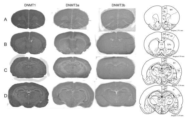
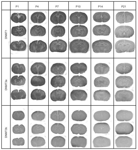
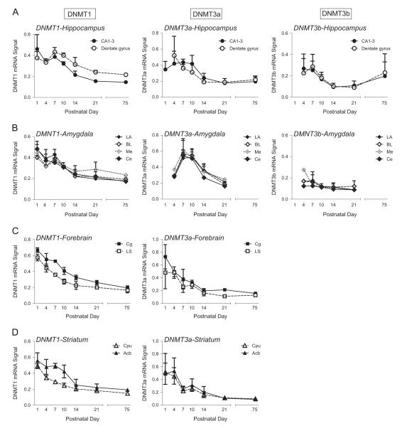
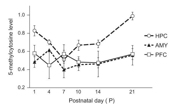
In spite of intense interest in how altered epigenetic processes including DNA methylation may contribute to psychiatric and neurodevelopmental disorders, there is a limited understanding of how methylation processes change during early postnatal brain development. The present study used in situ hybridization to assess mRNA expression for the three major DNA methyltranserases (DNMTs)--DNMT1, DNMT3a and DNMT3b--in the developing rat brain at seven developmental timepoints: postnatal days (P) 1, 4, 7, 10, 14, 21, and 75. We also assessed 5-methylcytosine levels (an indicator of global DNA methylation) in selected brain regions during the first three postnatal weeks. DNMT1, DNMT3a and DNMT3b mRNAs are widely expressed throughout the adult and postnatal developing rat brain. Overall, DNMT mRNA levels reached their highest point in the first week of life and gradually decreased over the first three postnatal weeks within the hippocampus, amygdala, striatum, cingulate and lateral septum. Global DNA methylation levels did not follow this developmental pattern; methylation levels gradually increased over the first three postnatal weeks in the hippocampus, and remained stable in the developing amygdala and prefrontal cortex. Our results contribute to a growing understanding of how DNA methylation markers unfold in the developing brain, and highlight how these developmental processes may differ within distinct brain regions.

Keywords: Amygdala; DNA methylation; DNA methyltransferase; Hippocampus; In situ hybridization; Neurodevelopment.


Figures

Fig. 1
Fig. 1
DNMT1, DNMT3a and DNMT3b mRNA expression in the adult rat brain. Columns show autoradiograms from representative tissue sections through the adult rat brain processed by in situ hybridization with antisense probes against rat DNMT1 (left), DNMT3a (middle) and DNMT3b (right) mRNA. X-ray films were exposed for 8 days for DNMT1, 3 days for DNMT3a, and 17 days for DNMT3b. Images show examples of coronal sections through the adult Sprague–Dawley rat brain, depicting regions such as the hippocampus, amygdala, caudate putamen, and hypothalamus. The diagrams in the far right column (adapted from Paxinos and Watson, 1997) indicate anatomical landmarks for each section. Abbreviations can be found in Table 1.
Fig. 2
Fig. 2
DNMT1, DNMT3a and DNMT3b mRNA expression in the developing rat brain. Columns show autoradiograms from representative tissue sections through the developmental rat brain at postnatal day (P)1, P4, P7, P10, P14, and P21 that were processed by in situ hybridization with antisense probe against rat DNMT1 (top column), DNMT3a (middle column) and DNMT3b (bottom column) mRNA. X-ray films were exposed for 8 days for DNMT1, 3 days for DNMT3a, and 17 days for DNMT3b.
Fig. 3
Fig. 3
Developmental patterns of DNMT1, DNMT3a and DNMT3b mRNA expression in the early rodent postnatal period and adulthood. DNA methyltransferase (DNMT)1, DNMT3a, and DNMT3b mRNA expression levels were quantified in several regions of rat brains collected at 6 developmental timepoints – postnatal day (P)1, P4, P7, P10, P14, P21, and P75 (adulthood). (A) Developmental expression pattern of DNMT1, DNMT3a, and DNTM3b in subregions of the hippocampus (CA1–3 and dentate gyrus). (B) Developmental expression pattern of DNMT1, DNMT3a, and DNMT3b in subnuclei of the amygdala (lateral, basolateral, medial and central nucleus). (C) Developmental expression pattern of DNMT1 and DNMT3a in several forebrain regions, including cingulate (Cg) and lateral septum (LS). Due to low abundance, DNMT3b was unable to be quantified in these regions. (D) Developmental expression pattern of DNMT1 and DNMT3a in the striatum (caudate putamen (Cpu) and nucleus accumbens (Acb)). Due to low abundance, DNMT3b was unable to be quantified in these regions.
Fig. 4
Fig. 4
Patterns of global DNA methylation in select brain regions of the developing rat brain. Global DNA methylation levels (approximated by measuring levels of 5-methylcytosine) were measured at several postnatal time periods – postnatal day (P)1, P4, P7, P10, P14 and P21 in tissue punches from the hippocampus, amygdala, and prefrontal cortex. Global DNA methylation levels appeared to increase, although this effect is most apparent in the developing hippocampus. Also, when comparing global DNA methylation levels across the three brain regions, levels were significantly higher in hippocampus compared to amygdala and prefrontal cortex.

References

    1. Agnish ND, Keller KA. The rationale for culling of rodent litters. Fundam. Appl. Toxicol.: Off. J. Soc. Toxicol. 1997;38:2–6. - PubMed
    1. Akbarian S. Epigenetics of schizophrenia. Curr. Top. Behav. Neurosci. 2010;4:611–628. - PubMed
    1. Beneyto M, Meador-Woodruff JH. Expression of transcripts encoding AMPA receptor subunits and associated postsynaptic proteins in the macaque brain. J. Comp. Neurol. 2004;468:530–554. - PubMed
    1. Broide RS, Redwine JM, Aftahi N, Young W, Bloom FE, Winrow CJ. Distribution of histone deacetylases 1–11 in the rat brain. J. Mol. Neurosci.: MN. 2007;31:47–58. - PubMed
    1. Brown SE, Weaver IC, Meaney MJ, Szyf M. Regional-specific global cytosine methylation and DNA methyltransferase expression in the adult rat hippocampus. Neurosci. Lett. 2008;440:49–53. - PubMed

Publication types

LinkOut - more resources