. 2013 Oct 3;13(4):471-82.
doi: 10.1016/j.stem.2013.07.010. Epub 2013 Aug 15.

The epidermis comprises autonomous compartments maintained by distinct stem cell populations

Affiliations

The epidermis comprises autonomous compartments maintained by distinct stem cell populations

Mahalia E Page et al. Cell Stem Cell. .

Abstract

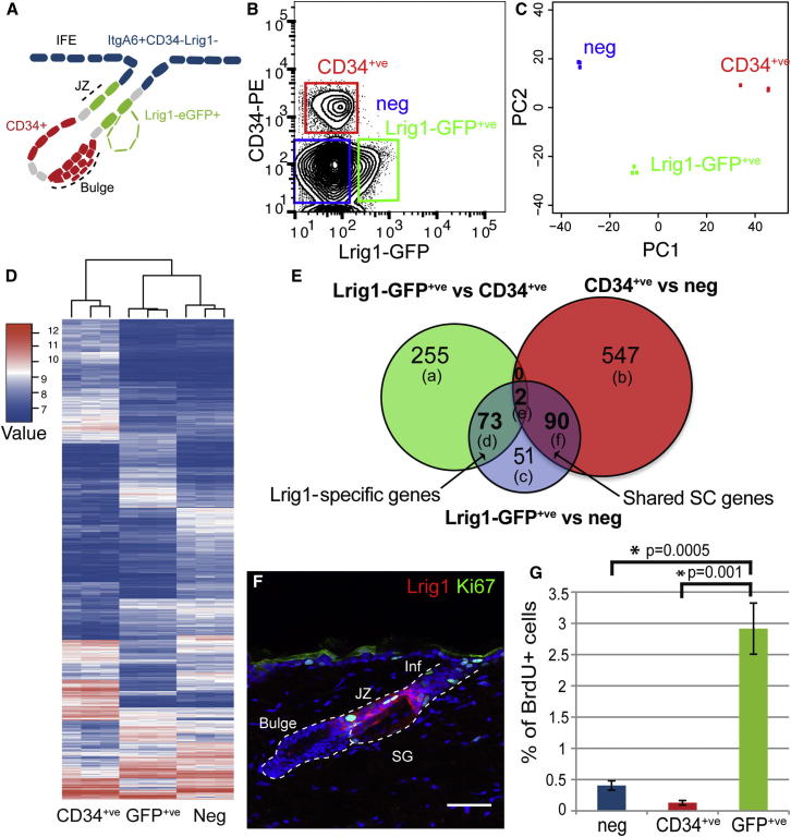
The complex anatomy of the epidermis contains multiple adult stem cell populations, but the extent to which they functionally overlap during homeostasis, wound healing, and tumor initiation remains poorly defined. Here, we demonstrate that Lrig1(+ve) cells are highly proliferative epidermal stem cells. Long-term clonal analysis reveals that Lrig1(+ve) cells maintain the upper pilosebaceous unit, containing the infundibulum and sebaceous gland as independent compartments, but contribute to neither the hair follicle nor the interfollicular epidermis, which are maintained by distinct stem cell populations. In contrast, upon wounding, stem cell progeny from multiple compartments acquire lineage plasticity and make permanent contributions to regenerating tissue. We further show that oncogene activation in Lrig1(+ve) cells drives hyperplasia but requires auxiliary stimuli for tumor formation. In summary, our data demonstrate that epidermal stem cells are lineage restricted during homeostasis and suggest that compartmentalization may constitute a conserved mechanism underlying epithelial tissue maintenance.


Figures

None
Graphical abstract
Figure 1
Figure 1
Characterization of SC Heterogeneity in the Epidermis (A) Schematic diagram of SC populations in the mouse pilosebaceous unit. Infund or Inf, infundibulum; Bulge, HF-SC niche. (B–D) Detection of Lrig1 protein (red) (B), Lrig1-EGFP (green) (C), and overlap (D) in telogen back skin. (E) Detection of Lrig1 (red) and CD34 (green). (F) Flow-cytometric analysis of Sca-1 expression in Lrig1-EGFP+ve cells (green line) from telogen back skin. The black line indicates basal epidermal cells based on ItgA6 expression. (G) Detection of Lrig1 (red) and Lgr5-EGFP (green) in anagen back skin. Is, isthmus; ORS, outer root sheath. (H and I) Detection of Lrig1 (red) and Plet-1 (green) (H) and Lgr6-EGFP (green) (I) in telogen back skin. Arrows indicate Lgr6-EGFP expression in the IFE. The arrowhead indicates Lgr6-EGFP expression in the SG. (J) Expression of Lrig1-EGFP (green) and Blimp1 (red) in the SG. (K) qPCR analysis of marker expression in Lrig1-EGFP- and CD34-expressing cells. Error bars represent the SEM (n = 4). Nuclei are counterstained with DAPI (blue). Scale bars represent 50 μm. See also Figure S1.
Figure 2
Figure 2
Lrig1 Marks a Unique and Highly Proliferative Compartment in the Epidermis (A) Schematic diagram of isolated cell populations. Bulge, HF-SC niche. (B) Isolation of ItgA6+veLrig1-EGFP+veCD34−ve (Lrig1-EGFP+ve, green) ItgA6+veLrig1-EGFP−veCD34+ve (CD34+ve, red) and ItgA6+veLrig1-EGFP−veCD34−ve (neg, blue) cells by flow cytometry. (C) Principal component analysis of gene-expression profiles associated with Lrig1-EGFP+ve, CD34+ve, and negative cells. (D and E) Heat map with the associated hierarchal clustering (D) and Venn diagram (E) of the upregulated probe sets associated with CD34+ve, Lrig1-GFP+ve, and negative epidermal cells. The number of individual probe sets in individual segments are indicated, and the associated probe sets are listed in Table S3 under (a)–(f). The (d) segment denotes genes enriched in Lrig1-EGFP+ve cells, and the (f) segment denotes genes shared by Lrig1-EGFP+ve and CD34+ve cells. (F) Detection of Ki67 (green) and Lrig1 (red) in telogen back skin. Nuclei are counterstained with DAPI (blue). Scale bars represent 50 μm. (G) Quantification of BrdU incorporation after a 1 hr chase by flow cytometry. Error bars represent the SEM (n = 4). See also Tables S1, S2, and S3.
Figure 3
Figure 3
The Epidermis Is Maintained in Discrete Compartments (A) Quantification of the proportion of labeled pilosebaceous units containing 1, 2, or >2 labeled cells 3 days (3d) post labeling. n = 100 pilosebaceous units (PSUs) from three mice. (B) Quantification of clone distribution at 3 days and 6 months (6m) post labeling. n = 100 pilosebaceous units from three mice. (C–J) Detection of tdTomato (red) and Lrig1-EGFP (green) after initiation of lineage tracing from Lrig1-expressing cells. Note the expansion of labeled progeny from Lrig1+ve cells into the infundibulum and in SG over time and that labeled cells do not contribute to the IFE (C–H). HG, hair germ. (K and L) Detection of tdTomato-labeled progeny of Lrig1-expressing cells and Blimp1 2 days and 3 months after labeling. Arrows indicate (K) initially labeled cells and (L) Blimp1+ve progeny. (M–P) Detection of tdTomato (red) after initiation of lineage tracing from Lgr5 (M and N) or K19 (O and P)-driven cre strains. Note that progeny of Lgr5+ve and K19+ve cells are restricted to the lower pilosebaceous unit. Nuclei are counterstained with DAPI (blue). Scale bars represent 50 μm. See also Figure S2.
Figure 4
Figure 4
Pilosebaceous SCs Break Compartment Boundaries in Response to Wounding and Convert to an IFE Fate (A–C and G–I) Serial fluorescence imaging of tdTomato-labeled progeny of Lrig1+ve (A–C) and Lgr5+ve (G–I) cells in the reepithelized wound (red). (D–F and J–L) Immunostaining of progeny of Lrig1+ve (D–F) or Lgr5+ve (J–L) cells in the regenerated IFE 10 months or 1 year after wounding. (M) Quantification of labeling after wounding of Lrig1, Lgr5, and K19 CreER mice by serial fluorescence imaging normalized to area of wound labeled 10 days after wounding. Error bars represent the SEM (Lgr5, n = 4; K19, n = 3; Lrig1, n = 5 mice). (N and O) Progeny of Lrig1+ve (N) and Lgr5+ve (O) cells (red) at day 2 following tail wounding (distance quantified in Figure S3F). (P–R) Progeny of Lrig1+ve cells detected in whole mounts of tail wound epidermis. The demarcated lines depict the wound margin. Nuclei are counterstained with DAPI (blue). Scale bars represent 1 mm (A–C and G–I), 50 μm (D–F and J–L), 100 μm (N and O), and 500 μm (P–R). See also Figure S3.
Figure 5
Figure 5
Wounding Induces K-Ras-Driven Papillomas (A) Spontaneous papilloma development in Lrig1 KI/LSL-K-Ras (G12D) mice (lip, n = 6; back, n = 8). (B) Lrig1-EGFP (green) detected in lip epidermis. (C) Detection of Lrig1 (red) and Ki67 (green) in lip epidermis. (D and E) Progeny of Lrig1+ve cells (red) detected in lip epidermis after short and long chase. Arrows indicate absence of labeling in the IFE adjacent to unlabeled infundibula. (F) TPA- and wound-induced papilloma development in Lrig1 KI/LSL-K-Ras (G12D) mice (TPA, n = 5; wound, n = 8). (G–M) Detection of tdTomato-labeled progeny of Lrig1-expressing cells in the IFE 4 days after the final TPA treatment of Lrig1 KI/LSL tdTomato mice. Hematoxylin and eosin (H&E) stain of lip papilloma (H) and (I) wound-induced papilloma on the back. Detection of Ki67 (green) and keratin 10 (green) in papillomas on the lip (J and K) and back (L and M) counterstained with keratin 14 (red) and DAPI (blue). Scale bars represent 50 μm (B and G), 100 μm (C–E), 1 mm (H), 5 mm (I), and 200 μm (J–M). See also Figure S4.

References

    1. Alcolea M.P., Jones P.H. Tracking cells in their native habitat: lineage tracing in epithelial neoplasia. Nat. Rev. Cancer. 2013;13:161–171. - PubMed
    1. Barker N., van Es J.H., Kuipers J., Kujala P., van den Born M., Cozijnsen M., Haegebarth A., Korving J., Begthel H., Peters P.J., Clevers H. Identification of stem cells in small intestine and colon by marker gene Lgr5. Nature. 2007;449:1003–1007. - PubMed
    1. Barker N., van Oudenaarden A., Clevers H. Identifying the stem cell of the intestinal crypt: strategies and pitfalls. Cell Stem Cell. 2012;11:452–460. - PubMed
    1. Blanpain C. Stem cells: Skin regeneration and repair. Nature. 2010;464:686–687. - PubMed
    1. Brownell I., Guevara E., Bai C.B., Loomis C.A., Joyner A.L. Nerve-derived sonic hedgehog defines a niche for hair follicle stem cells capable of becoming epidermal stem cells. Cell Stem Cell. 2011;8:552–565. - PMC - PubMed

Publication types