. 2013 Nov 12:252:126-43.
doi: 10.1016/j.neuroscience.2013.08.008. Epub 2013 Aug 15.

Dopamine D₂ and acetylcholine α7 nicotinic receptors have subcellular distributions favoring mediation of convergent signaling in the mouse ventral tegmental area

Affiliations

Dopamine D₂ and acetylcholine α7 nicotinic receptors have subcellular distributions favoring mediation of convergent signaling in the mouse ventral tegmental area

M Garzón et al. Neuroscience. .

Abstract

Alpha7 nicotinic acetylcholine receptors (α7nAChRs) mediate nicotine-induced burst-firing of dopamine neurons in the ventral tegmental area (VTA), a limbic brain region critically involved in reward and in dopamine D2 receptor (D2R)-related cortical dysfunctions associated with psychosis. The known presence of α7nAChRs and Gi-coupled D2Rs in dopamine neurons of the VTA suggests that these receptors are targeted to at least some of the same neurons in this brain region. To test this hypothesis, we used electron microscopic immunolabeling of antisera against peptide sequences of α7nACh and D2 receptors in the mouse VTA. Dual D2R and α7nAChR labeling was seen in many of the same somata (co-localization over 97%) and dendrites (co-localization over 49%), where immunoreactivity for each of the receptors was localized to endomembranes as well as to non-synaptic or synaptic plasma membranes often near excitatory-type synapses. In comparison with somata and dendrites, many more small axons and axon terminals were separately labeled for each of the receptors. Thus, single-labeled axon terminals were predominant for both α7nAChR (57.9%) and D2R (89.0%). The majority of the immunolabeled axonal profiles contained D2R-immunoreactivity (81.6%) and formed either symmetric or asymmetric synapses consistent with involvement in the release of both inhibitory and excitatory transmitters. Of 160 D2R-labeled terminals, 81.2% were presynaptic to dendrites that expressed α7nAChR alone or together with the D2R. Numerous glial processes inclusive of those enveloping either excitatory- or inhibitory-type synapses also contained single labeling for D2R (n=152) and α7nAChR (n=561). These results suggest that classic antipsychotic drugs, all of which block the D2R, may facilitate α7nAChR-mediated burst-firing by elimination of D2R-dependent inhibition in neurons expressing both receptors as well as by indirect pre-synaptic and glial mechanisms.

Keywords: ABC; ACh; Avidin–biotin complex; BSA; D(2)R; G-protein-coupled inwardly rectifying potassium; GIRK; MLA; N-methyl-d-aspartate receptor; NAc; NMDA; PB; PBS; TS; Tris-buffered saline; VTA; acetylcholine; addiction; alpha7 nicotinic acetylcholine receptors; bovine serum albumin; dopamine D(2) receptor; electron microscopic immunolabeling; mesocorticolimbic; methyllycaconitine; nAChR; nicotinic acetylcholine receptor; nucleus accumbens; phosphate buffer; phosphate-buffered saline; reward; schizophrenia; ventral tegmental area; α7nAChRs.


Figures

Fig. 1
Fig. 1
Photomicrographs showing immunoperoxidase labeling for the D2R in the caudate-putamen nucleus (CPu) of wild-type mice (A) is largely gone in the CPu of MSN-D2 KO mice (B). In contrast, there is no noticeable difference in the density of the labeling in the VTA of wild-type (C) and KO mice (D). Corner arrows point dorsal (d) and medial (m). Scale bars=0.02 μm.
Fig. 2
Fig. 2
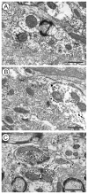
α7nAChR distribution in somata and dendrites with or without D2R. (A) A dark precipitous α7nAChR-immunoperoxidase reaction product (white block arrows) is observed rimming a multivesicular body (mvb) and the cisterns of endoplasmic reticulum and other endomembranes, as well as the plasma membrane of a neuronal soma (α7-soma). The soma receives synaptic contact from an axon terminal showing D2R gold particles (D2-t). (B) Aggregates of α7nAChR-immunoperoxidase (white block arrows) are observed within a dendrite receiving synaptic input from an unlabeled axon terminal (ut). (C) A clump of α7nAChR-immunoperoxidase reaction product (white block arrow) is localized in a dendrite (α7+D2-d) that also contains one cytoplasmic immunogold particle (small black arrow) for D2R. The dendrite receives an asymmetric synapse (black curved arrow) from an unlabeled axon terminal (ut). (D) Diffuse α7nAChR-immunoperoxidase labeling is seen in a dendrite (α7+D2-d) that also contains D2R-immunogold particles (small black arrows). The α7+D2-d shows an extensive plasmalemmal apposition with a dendrite exclusively labeled for D2R (D2-d), which receives what appears to be an asymmetric synapse from an unlabeled axon terminal (ut). (E) α7nAChR-immunoperoxidase aggregates (white block arrows) are clustered in an otherwise diffusely labeled dendrite (α7+D2-d) that also contains both cytoplasmic and plasmalemmal gold particles (small black arrow) for D2R. The dually-labeled α7+D2-d receives an asymmetric synapse (curved black arrow) from an unlabeled terminal (ut). (F) An unlabeled axon terminal (ut) makes a thick synaptic contact with a dendrite (α7+D2-d) showing D2R gold particles (small black arrows). The D2R immunogold particles are located beneath the postsynaptic specialization and distant from the synapse near an aggregate of α7nAChR-immunoperoxidase reaction product. Scale bars=500 nm.
Fig. 3
Fig. 3
D2R immunoperoxidase in axon terminals presynaptic to α7nAChR-labeled dendrites. (A) An axon terminal containing D2R immunoperoxidase labeling (D2-t) forms an asymmetric synapse with a dendrite (α7+D2-d) showing multiple cytoplasmic gold particles for α7nAChR (small black arrows) and also immunoperoxidase precipitate for D2R in tubulovesicular membranes (white block arrows). (B) A D2R-labeled terminal (D2-t) is presynaptic to dendrite showing plasmalemmal gold particles (small black arrows) for α7nAChR (α7-d) and receiving convergent synaptic input from an unlabeled axon terminal (ut). (C) D2R-immunoperoxidase labeling is seen in an axon terminal that forms an asymmetric synapse with a dendrite containing α7nAChR-immunogold particles (small black arrows) in the cytoplasm and in extrasynaptic and synaptic portions of the plasma membrane (α7-d). (D) D2R-immunperoxidase precipitate is seen within several axon terminals (D2-t) making contacts with dendrites showing immunogold particles (small black arrows) for α7nAChR (α7-d). In two of those terminals (D2-t1,2) showing discernible synapses with the dendrites, the D2R-immunoperoxidase is seen concentrated in the presynaptic sides facing α7-d1,2. The surrounding neuropil contains two small unmyelinated axons (α7-a1,2) with plasmalemmal α7nAChR immunogold particles (small black arrows. Scale bars=500 nm.
Fig. 4
Fig. 4
Subcellular distribution of D2R in axon terminals providing input to either D2R- or α7nAChR-immuolabeled dendrites. (A) D2R immunogold particles are seen within the cytoplasm and on small synaptic vesicles of an axon terminal (D2-t) that makes a symmetric synapse onto a dendrite (D2-d) containing also D2R-immunogold (small black arrows). (B) An axon terminal (D2-t) containing D2R-immunogold makes an asymmetric synapse with a dendrite (D2-d) showing synaptic D2R-immunogold particles (small black arrows). (C) D2R gold particles are observed rimming the membranes of small synaptic vesicles (ssv) within an axon terminal (D2-t) establishing an apparently asymmetric synapse with a dendrite (α7-d) containing a dense intracytoplasmic aggregate of α7nAChR-immunoperoxidase. (D) α7nAChR-immunoperoxidase clumps are seen beneath extrasynaptic portions of the plasma membrane of a dendrite (α7-d) receiving asymmetric synaptic input from a D2R immunogold-labeled terminal (D2-t1). The D2-t1 shows an appositional contact with another axon terminal (D2-t2) containing D2R gold particles (small black arrows). D2R immunogold=small black arrows; Scale bars=500 nm.
Fig. 5
Fig. 5
Co-localization of D2R and α7nAChR labeling in dendrites. (A) α7nAChR immunogold particles (small black arrows) are seen within the cytoplasm and on the postsynaptic density of an incoming synapse from an unlabeled terminal (ut) within a dendrite (α7+D2-d) that also contains diffuse D2R-immunoperoxidase labeling. (B) An axon terminal (D2-t) showing dense D2R immunoperoxidase makes what appears to be an obliquely sectioned asymmetric synapse with a dendrite (α7+D2-d) containing both light D2R immunoperoxidase and α7nAChR immunogold particles (small black arrows). (C) α7nAChR immunogold particles (small black arrows) and D2R immunoperoxidase are seen within a dendrite (α7+D2-d) that receives convergent input from two unlabeled axon terminals (ut1,2). A dendrite containing exclusively D2R immunoperoxidase labeling (D2-d) and a small unmyelinated axon (α7-a) showing plasmalemmal α7nAChR immunogold particles (small black arrow) are observed in the nearby neuropil. Scale bars=500 nm.
Fig. 6
Fig. 6
α7nAChR distribution in unmyelinated axons and glia. (A) α7nAChR immunoperoxidase is seen in transversally sectioned unmyelinated axons that show organized arrangement of microtubules (α7-a1–3). (B) Diffuse α7nAChR immunoperoxidase is observed in a small unmyelinated axon (α7-a) contacting a dendrite (α7+D2-d) showing α7nAChR immunoperoxidase and D2R immunogold (small black arrows). The α7nAChR immunoperoxidase is also observed near the plasma membrane of an astrocytic expansion (α7-g1). Another astrocytic process (α7-g2), lightly gray-shaded and depicting contour in discontinuous trace for easier recognition, contains aggregates of α7nAChR immunoperoxidase). The α7-g2 opposes and partially wraps the dually-labeled α7+D2-d. (C) Immunogold-labeled α7nAChR (small black arrows) is seen in a terminal (α7-t) making an asymmetric synapse (curved arrow) onto a α7-labeled dendrite (α7-d). A peroxidase-labeled D2R-labeled terminal is located adjacent to the same dendrite (curved white arrow). A small tubulovesicle (tv) is associated with two α7 gold particles. Unlabeled Dendrite=ud, α7 axon=α7-a. Scale bars=500 nm.
Fig. 7
Fig. 7
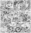
Glial distribution of α7nAChR, D2 receptor and S100. (A) An intensely α7nAChR-immunoperoxidase-labeled glial profile (α7-g) opposes a dendrite (α7+D2-d) containing D2R immunogold (small black arrows) and light α7nAChR-immunoperoxidase reaction product. The labeled glial process also partially contacts a D2R immunogold (small black arrows)-labeled axon terminal (D2-t) presynaptic to the α7+D2-d. (B) The α7nAChR-immunoperoxidase is seen in a glial profile (α7-g) opposing a dendrite (α7-d) that also contains clumps of α7nAChR peroxidase reaction product. The α7-g has thin expansions that partially form a common coating for α7-d and a D2R immunogold (small black arrows)-labeled axon terminal (D2-t) presynaptic to an unlabeled dendrite. (C) Glial profiles containing both α7nAChR gold particles (small black arrows) and immunoperoxidase D2R labeling (α7nAChR+D2R-g) are opposed to a dual-labeled α7nAChR+D2R-d. The dually labeled dendrite is receptive to an axon terminal containing peroxidase labeling for the D2R (D2-t). Insert shows a comparable dual-labeled glial process in another plane of section. (D and E) Immunogold D2R labeling (small arrows) is seen in glial profiles containing immunoperoxidase labeling for S100 (S100+D2-g). In D, the dually labeled glial process opposes many unlabeled axon terminals (ut) and a dendrite that also contains D2R immunogold (D2-d). In E, the dually labeled glial profile opposes many unlabeled dendritic and axonal profiles, while D2R (small arrows) is seen in a more distant dendrite (D2-d) and presynaptic terminal (D2-t). Scale bar=500 nm.
Fig. 8
Fig. 8
Bar graphs showing the synaptic area density (number of synapses per surface unit area) distribution of asymmetric or symmetric synapses made by axon terminals immunolabeled for D2R (D2R) and/or α7nAChR (α7R) with dendrites in the VTA. These dendrites are unlabeled (Un) or contain immunolabeling for α7R, D2R or both receptors (dual). Synaptic area density, shown in the Y axis, is expressed as the number of synapses per 1000 μm2 of analyzed tissue, calculated from the tallied micrograph area in which the synapses were counted for each group. The raw numbers of synapses are shown on top of each bar as the ratio value with respect to the total number of synapses within the group. Data were collected from six vibratome sections through the VTA in two mice. In three sections markers were reversed (immunogold or immunoperoxidase) to the other three, and both animals included sections of either type.

Similar articles

Cited by

References

    1. Ahn KC, Bernier BE, Harnett MT, Morikawa H. IP3 receptor sensitization during in vivo amphetamine experience enhances NMDA receptor plasticity in dopamine neurons of the ventral tegmental area. J Neurosci. 2010;30:6689–6699. - PMC - PubMed
    1. Amantea D, Bowery NG. Reduced inhibitory action of a GABAB receptor agonist on [3H]-dopamine release from rat ventral tegmental area in vitro after chronic nicotine administration. BMC Pharmacol. 2004;4:24. - PMC - PubMed
    1. Araque A, Parpura V, Sanzgiri R, Haydon P. Glutamate-dependent astrocyte modulation of synaptic transmission between cultured hippocampal neurons. Eur J Neurosci. 1998;10:2129–2142. - PubMed
    1. Arnaiz-Cot J, Gonzalez J, Sobrado M, Baldelli P, Carbone E, Gandia L, Garcia A, Hernandez-Guijo J. Allosteric modulation of alpha 7 nicotinic receptors selectively depolarizes hippocampal interneurons, enhancing spontaneous GABAergic transmission. Eur J Neurosci. 2008;27:1097–1110. - PubMed
    1. Aronin N, DiFiglia M. The subcellular localization of the G-protein Gi alpha in the basal ganglia reveals its potential role in both signal transduction and vesicle trafficking. J Neurosci. 1992;12:3435–3444. - PMC - PubMed

Publication types

MeSH terms

Substances

LinkOut - more resources