. 2013 Oct;12(10):1389-402.
doi: 10.1128/EC.00127-13. Epub 2013 Aug 16.

Impact of environmental conditions on the form and function of Candida albicans biofilms

Affiliations

Impact of environmental conditions on the form and function of Candida albicans biofilms

Karla J Daniels et al. Eukaryot Cell. 2013 Oct.

Abstract

Candida albicans, like other pathogens, can form complex biofilms on a variety of substrates. However, as the number of studies of gene regulation, architecture, and pathogenic traits of C. albicans biofilms has increased, so have differences in results. This suggests that depending upon the conditions employed, biofilms may vary widely, thus hampering attempts at a uniform description. Gene expression studies suggest that this may be the case. To explore this hypothesis further, we compared the architectures and traits of biofilms formed in RPMI 1640 and Spider media at 37°C in air. Biofilms formed by a/α cells in the two media differed to various degrees in cellular architecture, matrix deposition, penetrability by leukocytes, fluconazole susceptibility, and the facilitation of mating. Similar comparisons of a/a cells in the two media, however, were made difficult given that in air, although a/a cells form traditional biofilms in RPMI medium, they form polylayers composed primarily of yeast cells in Spider medium. These polylayers lack an upper hyphal/matrix region, are readily penetrated by leukocytes, are highly fluconazole susceptible, and do not facilitate mating. If, however, air is replaced with 20% CO2, a/a cells make a biofilm in Spider medium similar architecturally to that of a/α cells, which facilitates mating. A second, more cursory comparison is made between the disparate cellular architectures of a/a biofilms formed in air in RPMI and Lee's media. The results demonstrate that C. albicans forms very different types of biofilms depending upon the composition of the medium, level of CO2 in the atmosphere, and configuration of the MTL locus.


Figures
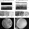
Fig 1
Fig 1
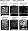
Biofilms formed by cells of a/α strain SC5314 in RPMI or Spider medium at 37°C in air exhibit differences in cellular architecture. (A and D) Comparison of side views of projection images of the stacked scans obtained by laser scanning confocal microscopy (LSCM) of a representative fixed, calcofluor white-stained biofilm formed in RPMI and Spider media, respectively. Orientation of hyphae can be assessed in the middle region. Intense calcofluor white staining at the top of each fixed biofilm is an artifact due to disproportionate binding after the biofilm is overlaid with the staining solution. (B and E) A comparison of individual LSCM scans through the lower, middle, and top portions of the alternative fixed, calcofluor white-stained representative a/α biofilms. The more vertical orientation of hyphae in the biofilm formed in RPMI medium is suggested in the middle scans by the white dots that represent cross sections of vertical hyphae. The less vertical orientation of hyphae in biofilms formed in Spider medium is suggested by the tangential sections of hyphae in the middle scans. Arrowheads indicate small clumps of cells. (C and F) A low-magnification comparison of an entire paraformaldehyde-fixed a/α biofilm formed in RPMI and Spider media, respectively. Notice the wrinkled membranous appearance of the former and the hairy, unwrinkled appearance of the latter. Low-magnification images were obtained through a Wild M8 stereo zoom microscope equipped with a Nikon Cool Pix camera. Note that highly similar results were obtained for the a/α strain P37037 (a/α) (data not shown).
Fig 2
Fig 2
Models of a/α and a/a biofilm formed in RPMI and Spider media at 37°C in air and the radical difference between a/a biofilms formed in Spider medium in air and those formed in 20% CO2. (A through E) The models were deduced from LSCM scans of five biofilms for each of two independent a/α strains and each of two independent a/a strains. Mixed MTL-homozygous white a/a-α/α (50:50) biofilms exhibited the same differences in architecture as did a/a biofilms in air versus 20% CO2 (data not shown). (F) Key for interpreting the models.
Fig 3
Fig 3
Biofilms formed by a/a cells in strain P37005 in RPMI and Spider media at 37°C in air exhibit dramatic differences in cellular architecture. While a/a cells form a biofilm in RPMI medium in air similar to that formed by a/α cells, a/a cells form a polylayer made up primarily of yeast cells lacking an upper region of hyphae and matrix in Spider medium in air, a phenotype quite distinct from a/α cells in Spider medium. (A and D) A comparison of side views of projection images of stacked LSCM scans. (B and E) A comparison of individual LSCM scans through the lower, middle, and top portions of the alternative fixed, calcofluor white-stained biofilm formed at 37°C in air in RPMI medium and the predominantly yeast cell polylayer formed at 37°C in air in Spider medium, respectively. The vertical orientation of hyphae is suggested by the white dots representing cross sections of hyphae in the middle-portion scans of the biofilm formed in RPMI medium in air, while the compact yeast cell nature of the polylayer formed by a/a cells in Spider medium in air is apparent in the lower, middle, and top scans. (C and F) A low-magnification comparison of the entire paraformaldehyde-fixed biofilms formed by a/a cells in RPMI and Spider media, respectively. Notice the wrinkled membranous nature of the a/a biofilm (C) formed in RPMI medium in air, similar to that formed by a/α cells in RPMI medium in air (compare with Fig. 1C). Notice the granular pattern of the a/a biofilm formed in Spider medium in air, reflecting the predominant yeast cell nature. The blow-up panel of cells in a space in the a/a polylayer formed in Spider medium in air reveals cells only in the budding yeast phase.
Fig 4
Fig 4
Staining with Sypro Ruby, a dye that stains both cell wall and matrix, reveals a denser matrix in the areas between the hyphae in an a/α biofilm (SC5314) formed in RPMI medium at 37°C in air (A) than in an a/α biofilm formed in Spider medium at 37°C in air (B). All images were obtained at the same LSCM settings.
Fig 5
Fig 5
Leukocyte penetrability and fluconazole susceptibility of a/α and a/a biofilms formed in RPMI medium versus Spider medium in air at 37°C. (A through D) Side views of the penetrability of biofilms by leukocytes. GFP-expressing leukocytes of the cell line HL60 were layered on top of mature 48-h biofilms and incubated for 3 h at 37°C in 5% CO2. The biofilms were then analyzed by LSCM. Side views of projection images are presented. The dashed lines represent the top of each biofilm. (E) Fluconazole susceptibility. Susceptibility was assessed by measuring the percent cells that stained with Sytox Green, a dead-cell double-stranded DNA stain. Total cells were measured by staining total nuclei with Hoechst 33342. Mature, 48-h biofilms or yeast cell polylayers were incubated with fluconazole for 24 subsequent hours before analysis.
Fig 6
Fig 6
While a/a cells form a thick biofilm with an upper hyphal/matrix region in Spider medium in 20% CO2, they form a much thinner, predominantly yeast cell polylayer, approximately 10 cells thick, in Spider medium in air. (A and C) Comparisons of top view (highest LSCM scan) of the alternative calcofluor white-stained a/a biofilms formed in air and 20% CO2, respectively. The former is comprised of yeast cells and the latter of hyphae. (B and D) Orthogonal 90° view of a single z-slice through LSCM projected scans in air and 20% CO2, respectively. The means and standard derivations of thickness in micrometers are presented below panel B and D.
Fig 7
Fig 7
Biofilms formed in air in RPMI medium according to the methods of Soll and colleagues (, , , –42, 44, 46, 47) versus those formed in Lee's medium according to the methods of Bennett and colleagues (24) differ dramatically in thickness and architecture. Those formed in RPMI medium are thick and consist of a basal yeast cell layer and upper hyphal/matrix region. Those formed in Lee's medium are thin and consist of a predominantly yeast cell polylayer on silicone elastomer or plastic. Pheromone treatment increases thickness and, notably, attachment to the plastic surface but does not alter the general differences in biofilm architecture. All preparations were stained with calcofluor white. (A, D, and G) Biofilm surface obtained by LSCM scans of untreated RPMI medium-derived biofilms on silicone elastomer and Lee's medium-derived biofilms on silicone elastomer or plastic, respectively. (B, E, and H) Side views of stacks of scans of the respective biofilms. (C, F, and I) Thickness stacks of LSCM scans of the respective, untreated biofilms. (J, M, and P) Biofilm surface obtained by LSCM scans of pheromone-treated RPMI medium-derived biofilms of silicone elastomer and Lee's medium-derived biofilms on silicone elastomer or plastic, respectively. (K, N, and Q) Orthogonal 90° view of a single z-slice through LSCM projected scans of respective pheromone-treated biofilms. (L, O, and R) Thickness and filamentation of respective pheromone-treated biofilms. Biofilm surface images at higher magnification can be seen in Fig. S1 in the supplemental material. Note that because preparations on silicone elastomer were viewed with an inverted microscope (20× objective) and those on plastic were viewed with an upright microscope (63× objective), magnification of the two differs by 4-fold (see scale bars).

Similar articles

Cited by

References

    1. Costerton JW, Stewart PS, Greenberg EP. 1999. Bacterial biofilms: a common cause of persistent infections. Science 284:1318–1322 - PubMed
    1. Hall-Stoodley L, Stoodley P. 2005. Biofilm formation and dispersal and the transmission of human pathogens. Trends Microbiol. 13:7–10 - PubMed
    1. Douglas LJ. 2003. Candida biofilms and their role in infection. Trends Microbiol. 11:30–36 - PubMed
    1. Finkel JS, Mitchell AP. 2011. Genetic control of Candida albicans biofilm development. Nat. Rev. Microbiol. 9:109–118 - PMC - PubMed
    1. Ramage G, Saville SP, Thomas DP, Lopez-Ribot JL. 2005. Candida biofilms: an update. Eukaryot. Cell 4:633–638 - PMC - PubMed

Publication types

MeSH terms

LinkOut - more resources