. 2013 Oct;31(10):916-21.
doi: 10.1038/nbt.2672. Epub 2013 Aug 18.

Nature-inspired design of motif-specific antibody scaffolds

Affiliations

Nature-inspired design of motif-specific antibody scaffolds

James T Koerber et al. Nat Biotechnol. 2013 Oct.

Abstract

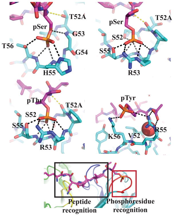
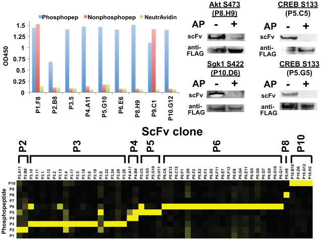
Aberrant changes in post-translational modifications (PTMs) such as phosphate groups underlie a majority of human diseases. However, detection and quantification of PTMs for diagnostic or biomarker applications often require PTM-specific monoclonal antibodies (mAbs), which are challenging to generate using traditional antibody-selection methods. Here we outline a general strategy for producing synthetic, PTM-specific mAbs by engineering a motif-specific 'hot spot' into an antibody scaffold. Inspired by a natural phosphate-binding motif, we designed and selected mAb scaffolds with hot spots specific for phosphoserine, phosphothreonine or phosphotyrosine. Crystal structures of the phospho-specific mAbs revealed two distinct modes of phosphoresidue recognition. Our data suggest that each hot spot functions independently of the surrounding scaffold, as phage display antibody libraries using these scaffolds yielded >50 phospho- and target-specific mAbs against 70% of target peptides. Our motif-specific scaffold strategy may provide a general solution for rapid, robust development of anti-PTM mAbs for signaling, diagnostic and therapeutic applications.


Figures

Figure 1
Figure 1
Design of phospho-specific Ab scaffold. a) Structure of CDR H2 loop from Ab (PDB ID 1i8i) bound to aspartate in peptide antigen. Each H2 residue contributes to anchoring the peptide (52H and 52AH), specificity (53H, 55H, and 56H), or conformation (53H). Hydrogen bonds that confer specificity are shown in black and anchoring hydrogen bonds are shown in yellow. The peptide is shown in magenta and Ab heavy chain is shown in cyan. b) Competition phage ELISAs with humanized Fab. Eight different mutant peptides containing D, A, S, T, Y, pS, pT, or pY at position 8 of the peptide were used as soluble competitors to inhibit Fab-phage binding to the immobilized wild-type peptide (KGNYVVTDH) (n=3, error bars represent standard deviation). Strong competition was observed for the wild-type peptide (green line), whereas no competition was observed for the S, T, A, or Y peptides (dashed lines) indicating that D is a hot-spot residue. Notably, the Fab binds to phosphorylated species as weak competition was observed for the pSer and pThr peptides (orange and blue solid lines, respectively). c) Representative pooled phage ELISAs from selection of H2-targeted library against pSer peptide. After three rounds of selection, all library pools exhibited higher binding signal to the pSer peptide than the parent Fab (dashed line).
Figure 2
Figure 2
Selection and characterization of pSer-, pSer/pThr-, and pTyr-specific scaffolds. Competition ELISAs were used to determine the specificity of each Ab scaffold (n=3, error bars represent standard deviation). For both pSAb (a) and pSTAb (b), no binding inhibition was observed for the unphosphorylated peptides up to 2 μM, whereas strong inhibition was observed for the phosphorylated peptides. For pYAb (c), weak inhibition was observed at high concentrations of the unphosphorylated Tyr peptide, but ~20-fold less pTyr peptide was required to observe the same level of inhibition. The sequence frequency logos of the Ab pools from which each lead clone was derived are depicted in the bottom panels. GS and H2 indicate the sequence logos from GS and H2 libraries selected against pSer and pThr. For the six-residue loops selected for pSer or pThr binding, clear enrichment for the G53H and G54H is seen. For the seven-residue loops selected for pSer or pThr binding, we observed a replacement of G53H with Pro-Arg, likely opening up the binding pocket to better accommodate pThr. All clones that bound pTyr came from the six-residue libraries and contain two positively charged amino acids at H55 and H56. The H2 sequences of pSAb, pSTAb, and pYAb are ATGGHT, STPRGST, and VTGGRK, respectively.
Figure 3
Figure 3
X-ray crystal structures of phosphoresidue-binding pocket from pSAb (a), pSTAb (b and c), and pYAb (d). a) In pSAb, pSer makes hydrogen bonds with all three specificity residues (G53H, H55H and T56H). The anchoring hydrogen bond (yellow) to T52AH is conserved. b and c) In pSTAb, the pSer/pThr makes hydrogen bonds with two specificity residues (R53H and S55H), one anchor residue (S52H), and the conformation residue (G54H). In both pSTAb structures bound to pSer and pThr, R53H forms a bidentate interaction with the phosphate. The anchor residue T52AH is flipped compared to pSAb, which allows the backbone carbonyl to make a new anchoring hydrogen bond (yellow). d) The pTyr is recognized by a salt bridge with K56H and a hydrophobic interaction between V52H and the phenyl ring of the pTyr. However, the phosphate group of pTyr does not occupy the phosphate-binding pocket, which is instead occupied by a water molecule (shown as red sphere). e) The structures demonstrate two distinct recognition sectors: a phosphoresidue-binding pocket (red box) and the peptide-binding “reader” region (black box). Key CDRs L3, H2, and H3 are colored yellow, dark blue, and red. Phosphopeptides are shaded magenta and the Ab light and heavy chains are shaded green and cyan, respectively. Yellow and black dashed lines indicate hydrogen bonds between the phosphoresidue and Ab scaffold.
Figure 4
Figure 4
Generation of recombinant phospho-specific (PS) Abs using the pSAb and pSTAb scaffolds. a) Representative phage ELISAs of one scFv clone selected against each of the nine phosphopeptide targets demonstrates that we selected PS Abs to seven out of the ten targets. No hits were observed against P7. To analyze target specificity, we characterized the binding of each scFv-phage to ten different phosphopeptides by phage ELISA (n = 2 – 3) (b). Heatmap representation of the phage ELISA binding signals for each scFv-phage (vertical axis) against each of the ten phosphopeptides (horizontal axis). Strikingly, most of these scFvs bind only to the phosphopeptide against which they were selected. For each scFv, signals were normalized to the highest overall ELISA signal observed against the ten peptides. The scale goes from zero (black) to one (yellow). c) ScFvs also recognize the phosphorylated protein in Western blots. FLAG-tagged target proteins were immunoprecipitated from transiently transfected HEK293T. To verify PS binding, samples were either dephosphorylated using alkaline phosphatase (AP) or treated with buffer only. Membranes were probed with biotinylated scFv (20 μg/mL) overnight and bound scFv was detected using NeutrAvidin-HRP. Total levels of target protein were monitored using anti-FLAG-HRP (Supplemental Methods).

Comment in

References

    1. Cohen P. The regulation of protein function by multisite phosphorylation--a 25 year update. Trends Biochem Sci. 2000;25:596–601. - PubMed
    1. Hanahan D, Weinberg RA. Hallmarks of cancer: the next generation. Cell. 2011;144:646–674. - PubMed
    1. Blagoev B, Ong SE, Kratchmarova I, Mann M. Temporal analysis of phosphotyrosine-dependent signaling networks by quantitative proteomics. Nat Biotechnol. 2004;22:1139–1145. - PubMed
    1. Zhou H, Watts JD, Aebersold R. A systematic approach to the analysis of protein phosphorylation. Nat Biotechnol. 2001;19:375–378. - PubMed
    1. Hornbeck PV, Chabra I, Kornhauser JM, Skrzypek E, Zhang B. PhosphoSite: A bioinformatics resource dedicated to physiological protein phosphorylation. Proteomics. 2004;4:1551–1561. - PubMed

Publication types