

doi: 10.1038/bcj.2013.33.
A single-tube multiparameter seven-colour flow cytometry strategy for the detection of malignant plasma cells in multiple myeloma
Affiliations
- PMID: 23955589
- PMCID: PMC3763387
- DOI: 10.1038/bcj.2013.33
Item in Clipboard
A single-tube multiparameter seven-colour flow cytometry strategy for the detection of malignant plasma cells in multiple myeloma
Blood Cancer J.
.
No abstract available
Figures
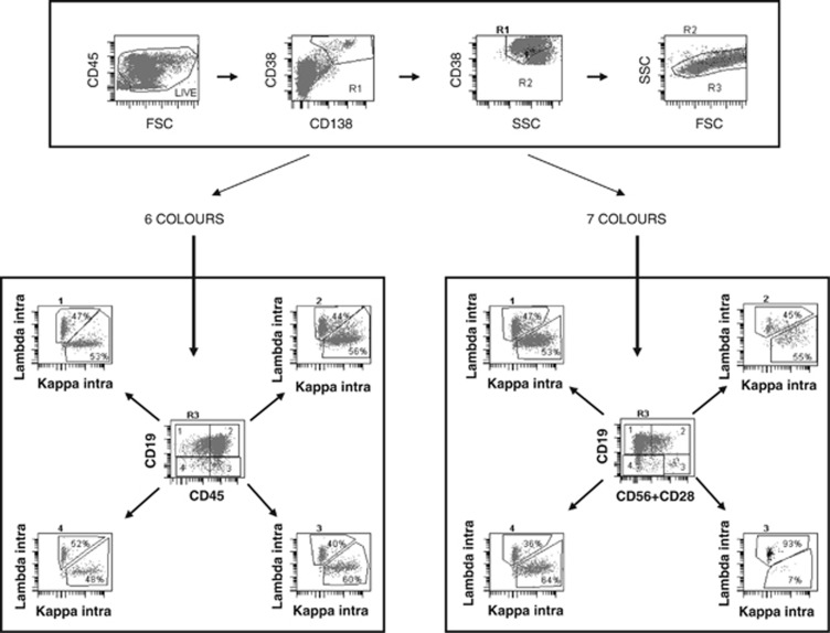
Analysis of the data from a single-tube seven-colour staining with a six-colour strategy (left) or a seven-colour strategy (right). After defining a nucleated cell gate on a FSC/SSC scattergram and excluding debris on an FSC/CD45 gate, PCs are first included in a broad R1 gate encompassing CD138+/CD38++ or CD38− cells. This population is refined on an SSC/CD38 scattergram conditioned on R1, thereby defining gate R2. Cells satisfying both R1 and R2 are then displayed on an FSC/SSC scattergram and included in an R3 gate. For six-colour analysis (left bottom panel), cells in R3 are displayed on a CD19/CD45 scattergram, allowing to define four populations as shown in the centre of the panel. For each of them, a κ/λ scattergram is established to discriminate normal polyclonal PCs and MM-restricted PCs. The right bottom panel shows that, in the seven-colour strategy, intracytoplasmic light chain restriction is examined among four different populations, delineated on the basis of the expression or not of CD19 combined to the mixture of CD28 and CD56 (either or both antigens expressed when positive). CD45, which can be abnormally expressed on the clonal population as shown in the six-colour strategy, is examined on a different plot. In this sample (same list mode), the six-colour strategy fails to identify light chain restriction (MRD <0.24 × 10⁻⁴), whereas the seven-colour strategy reveals that 93% of the cells in subset 3 (CD19−/CD56/CD28+) use lambda chains, thereby characterizing abnormal PCs (MRD 1.25 × 10⁻⁴).
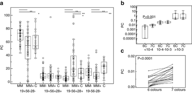
(a) Partition of plasma cells within the four subsets defined in seven-colour staining, based on the expression or not of CD19 and/or CD28/CD56 within samples with no MRD (MM−), positive MRD (MM+) or from normal controls (C). Statistically significant differences are indicated by asterisks: **<0.0001, *<0.01. (b) Comparison of MRD results for the same listmodes interpreted with a six (6C)- or a seven (7C)-colour strategy. Data do not differ for samples with more than 1 × 10⁻⁴ cells, but the seven-colour strategy is more efficient to detect the lowest levels (**<0.0001). (c) Paired comparison of the 17 samples with positive MRD below 10−4 in the seven-colour strategy analysed in six (left) or seven (right) colours.
References
-
- Rawstron AC, Davies FE, Das Gupta R, Ashcroft AJ, Patmore R, Drayson MT, et al. Flow cytometric disease monitoring in multiple myeloma: the relationship between normal and neoplastic plasma cells predicts outcome after transplantation. Blood. 2002;100:3095–3100. - PubMed
-
- Pellat-Deceunynck C, Bataille R, Robillard N, Harousseau JL, Rapp MJ, Juge-Morineau N, et al. Expression of CD28 and CD40 in human myeloma cells: a comparative study with normal plasma cells. Blood. 1994;84:2597–2603. - PubMed
-
- Robillard N, Wuillème S, Lodé L, Magrangeas F, Minvielle S, Avet-Loiseau H. CD33 is expressed on plasma cells of a significant number of myeloma patients, and may represent a therapeutic target. Leukemia. 2005;19:2021–2022. - PubMed
-
- Ocqueteau M, Orfao A, Garcia-Sanz R, Almeida J, Gonzalez M, San Miguel JF. Expression of the CD117 antigen (c-Kit) on normal and myelomatous plasma cells. Br J Haematol. 1996;95:489–493. - PubMed
LinkOut - more resources
Full Text Sources
Other Literature Sources
