Common variation at 3q26.2, 6p21.33, 17p11.2 and 22q13.1 influences multiple myeloma risk
- PMID: 23955597
- PMCID: PMC5053356
- DOI: 10.1038/ng.2733
Common variation at 3q26.2, 6p21.33, 17p11.2 and 22q13.1 influences multiple myeloma risk
Abstract
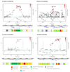
To identify variants for multiple myeloma risk, we conducted a genome-wide association study with validation in additional series totaling 4,692 individuals with multiple myeloma (cases) and 10,990 controls. We identified four risk loci at 3q26.2 (rs10936599, P = 8.70 × 10(-14)), 6p21.33 (rs2285803, PSORS1C2, P = 9.67 × 10(-11)), 17p11.2 (rs4273077, TNFRSF13B, P = 7.67 × 10(-9)) and 22q13.1 (rs877529, CBX7, P = 7.63 × 10(-16)). These data provide further evidence for genetic susceptibility to this B-cell hematological malignancy, as well as insight into the biological basis of predisposition.
Conflict of interest statement
Statement The authors declare no competing financial interests.
Figures

References
-
- Kyle RA, Rajkumar SV. Multiple myeloma. N Engl J Med. 2004;351:1860–73. - PubMed
-
- Palumbo A, Anderson K. Multiple myeloma. N Engl J Med. 2011;364:1046–60. - PubMed
-
- Schmermund A, et al. Assessment of clinically silent atherosclerotic disease and established and novel risk factors for predicting myocardial infarction and cardiac death in healthy middle-aged subjects: rationale and design of the Heinz Nixdorf RECALL Study. Risk Factors, Evaluation of Coronary Calcium and Lifestyle. Am Heart J. 2002;144:212–8. - PubMed
Publication types
MeSH terms
Grants and funding
LinkOut - more resources
Full Text Sources
Other Literature Sources
Medical

