

Optical coherence tomography imaging during thyroid and parathyroid surgery: a novel system of tissue identification and differentiation to obviate tissue resection and frozen section
- PMID: 23956009
- PMCID: PMC5777931
- DOI: 10.1002/hed.23452
Optical coherence tomography imaging during thyroid and parathyroid surgery: a novel system of tissue identification and differentiation to obviate tissue resection and frozen section
Abstract
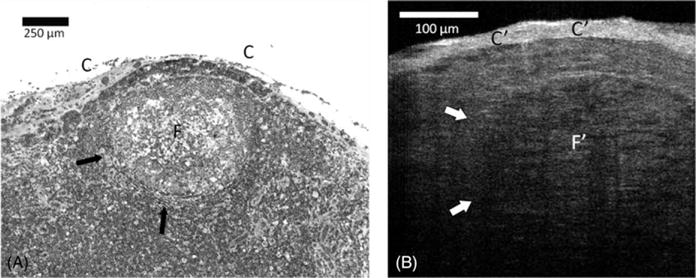
Background: Optical coherence tomography (OCT) allows tissue histologic-like evaluation, but without tissue fixation or staining. We investigated OCT images from tissues obtained at thyroid and parathyroid surgeries to provide a preliminary assessment as to whether these images contain sufficient information for recognition and differentiation of normal neck tissues.
Methods: Normal tissues were obtained from patients undergoing surgical treatment. Two new-generation OCT systems, including optical frequency domain imaging (OFDI) and μOCT, were compared to representative hematoxylin-eosin histology.
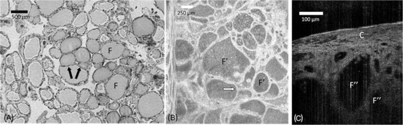
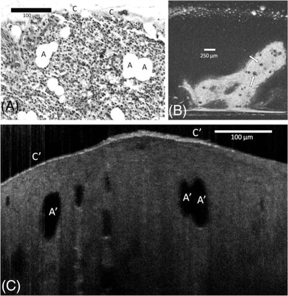
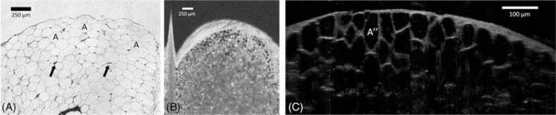
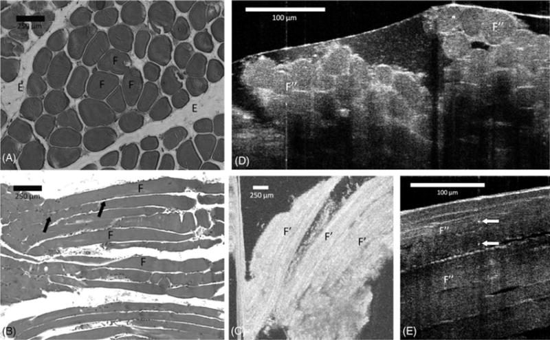
Results: Thyroid, fat, muscle, lymph nodes, and parathyroid tissues were evaluated. Histologic-like microscopic characteristics sufficient for tissue type identification was realized using both systems for all tissue types examined.
Conclusion: This pilot study demonstrated that new-generation OCT systems are capable of recognizing and differentiating neck tissues encountered during thyroid and parathyroid surgeries. Further advances in OCT miniaturization and development of sterile intraoperative probe formats may allow OCT to offer an intraoperative "optical biopsy" without fixation, staining, or tissue resection.
Keywords: hyperparathyroidism; parathyroid adenoma; parathyroidectomy; thyroid cancer; thyroidectomy.
Copyright © 2013 Wiley Periodicals, Inc.
Figures
Similar articles
-
Rapid head and neck tissue identification in thyroid and parathyroid surgery using optical coherence tomography.Head Neck. 2019 Dec;41(12):4171-4180. doi: 10.1002/hed.25972. Epub 2019 Oct 1. Head Neck. 2019. PMID: 31571306
-
Intraoperative use of optical coherence tomography to differentiate normal and diseased thyroid and parathyroid tissues from lymph node and fat.Lasers Med Sci. 2021 Mar;36(2):269-278. doi: 10.1007/s10103-020-03024-z. Epub 2020 Apr 27. Lasers Med Sci. 2021. PMID: 32337680 Free PMC article.
-
Intraoperative optical coherence tomography imaging to identify parathyroid glands.Surg Endosc. 2015 Sep;29(9):2698-704. doi: 10.1007/s00464-014-3992-x. Epub 2014 Dec 5. Surg Endosc. 2015. PMID: 25475518
-
Impact of fluorescence and autofluorescence on surgical strategy in benign and malignant neck endocrine diseases.Best Pract Res Clin Endocrinol Metab. 2019 Aug;33(4):101311. doi: 10.1016/j.beem.2019.101311. Epub 2019 Aug 10. Best Pract Res Clin Endocrinol Metab. 2019. PMID: 31494052 Review.
-
Thyroid and parathyroid surgeon case volume influences patient outcomes: A systematic review.Surg Oncol. 2021 Sep;38:101550. doi: 10.1016/j.suronc.2021.101550. Epub 2021 Apr 6. Surg Oncol. 2021. PMID: 33915486
Cited by
-
Carbon nanoparticles guide contralateral central neck dissection in patients with papillary thyroid cancer.Oncol Lett. 2018 Jul;16(1):447-452. doi: 10.3892/ol.2018.8691. Epub 2018 May 10. Oncol Lett. 2018. PMID: 29963128 Free PMC article.
-
Potential role of carbon nanoparticles in protection of parathyroid glands in patients with papillary thyroid cancer.Medicine (Baltimore). 2016 Oct;95(42):e5002. doi: 10.1097/MD.0000000000005002. Medicine (Baltimore). 2016. PMID: 27759629 Free PMC article. Clinical Trial.
-
In-vivo handheld optoacoustic tomography of the human thyroid.Photoacoustics. 2016 Jun 27;4(2):65-69. doi: 10.1016/j.pacs.2016.05.003. eCollection 2016 Jun. Photoacoustics. 2016. PMID: 27766210 Free PMC article.
-
[Optical coherence tomography for differentiation of parathyroid gland tissue].Chirurg. 2016 May;87(5):416-22. doi: 10.1007/s00104-015-0120-y. Chirurg. 2016. PMID: 26661948 German.
-
Wide-field optical coherence tomography for microstructural analysis of key tissue types: a proof-of-concept evaluation.Pathol Oncol Res. 2023 Jul 14;29:1611167. doi: 10.3389/pore.2023.1611167. eCollection 2023. Pathol Oncol Res. 2023. PMID: 37521364 Free PMC article.
References
-
- Mangano JJ. Geographic variation in U.S. thyroid cancer incidence and a cluster near nuclear reactors in New Jersey, New York, and Pennsylvania. Int J Health Serv. 2009;39:643–661. - PubMed
-
- Menegaux F, Turpin G, Dahman M, et al. Secondary thyroidectomy in patients with prior thyroid surgery for benign disease: a study of 203 cases. Surgery. 1999;126:479–483. - PubMed
-
- Khan AA, Bilezikian JP, Potts JT, Jr, Guest Editors for the Third International Workshop on Asymptomatic Primary Hyperparathyroidism The diagnosis and management of asymptomatic primary hyperparathyroidism revisited. J Clin Endocrinol Metab. 2009;94:333–334. - PubMed
-
- Eastell R, Arnold A, Brandi ML, et al. Diagnosis of asymptomatic primary hyperparathyroidism: proceedings of the third international workshop. J Clin Endocrinol Metab. 2009;94:340–350. - PubMed
-
- Uhlig K, Berns JS, Kestenbaum B, et al. KDOQI US commentary on the 2009 KDIGO clinical practice guideline for the diagnosis, evaluation, and treatment of CKD-mineral and bone disorder (CKD-MBD) Am J Kidney Dis. 2010;55:773–799. - PubMed
Publication types
MeSH terms
Grants and funding
LinkOut - more resources
Full Text Sources
Other Literature Sources
Medical
