. 2013:2013:231351.
doi: 10.1155/2013/231351. Epub 2013 Jul 17.

Lipoxin a4 preconditioning and postconditioning protect myocardial ischemia/reperfusion injury in rats

Affiliations

Lipoxin a4 preconditioning and postconditioning protect myocardial ischemia/reperfusion injury in rats

Qifeng Zhao et al. Mediators Inflamm. 2013.

Abstract

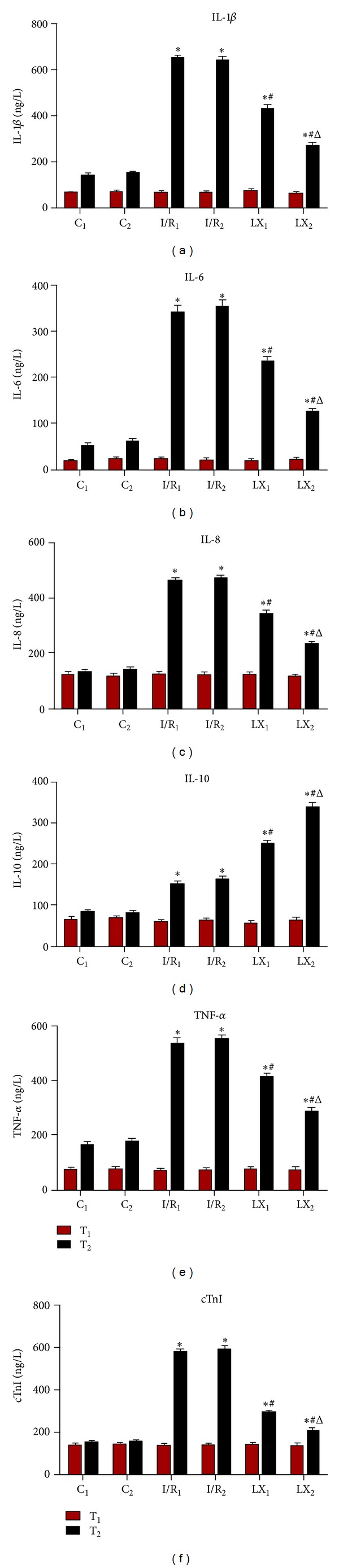
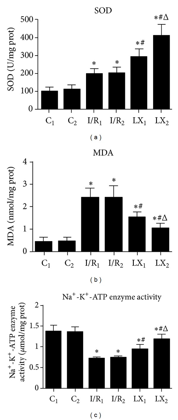
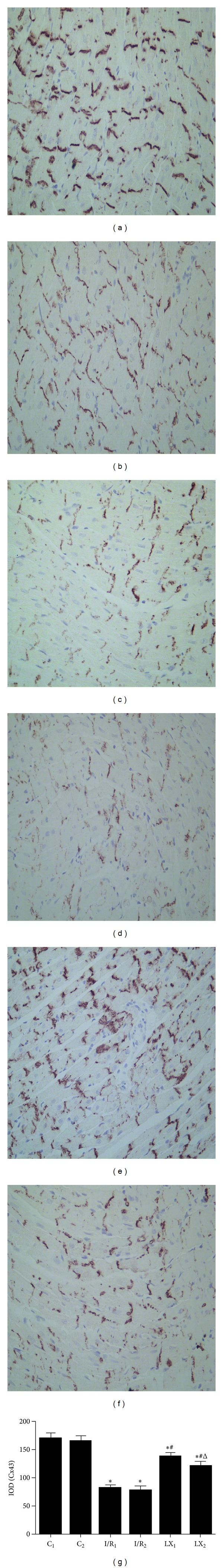
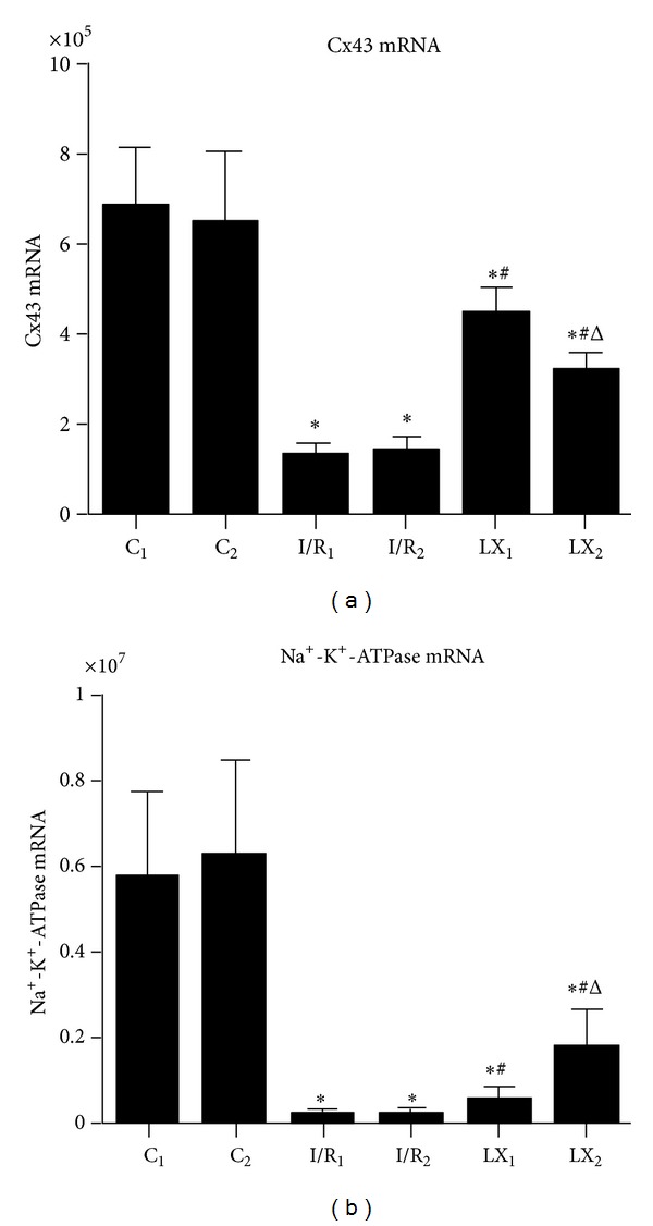
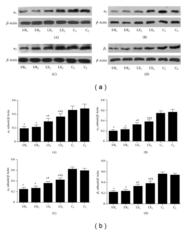
This study aims to investigate the pre- and postconditioning effects of lipoxin A4 (LXA4) on myocardial damage caused by ischemia/reperfusion (I/R) injury. Seventy-two rats were divided into 6 groups: sham groups (C1 and C2), I/R groups (I/R1 and I/R2), and I/R plus LXA4 preconditioning and postconditioning groups (LX1 and LX2). The serum levels of IL-1 β , IL-6, IL-8, IL-10, TNF- α , and cardiac troponin I (cTnI) were measured. The content and the activity of Na(+)-K(+)-ATPase as well as the superoxide dismutase (SOD), and malondialdehyde (MDA) levels were determined. Along with the examination of myocardium ultrastructure and ventricular arrhythmia scores (VAS), connexin 43 (Cx43) expression were also detected. Lower levels of IL-1 β , IL-6, IL-8, TNF- α , cTnI, MDA content, and VAS and higher levels of IL-10, SOD activity, Na(+)-K(+)-ATPase content and activity, and Cx43 expression appeared in LX groups than I/R groups. Besides, H&E staining, TEM examination as well as analysis of gene, and protein confirmed that LXA4 preconditioning was more effective than postconditioning in preventing arrhythmogenesis via the upregulation of Cx43. That is, LXA4 postconditioning had better protective effect on Na(+)-K(+)-ATPase and myocardial ultrastructure.


Figures

Figure 1
Figure 1
Comparison of concentrations of serum IL-1β, IL-6, IL-8, IL-10; TNF-α;, and cTnI at T1 and T2 among all groups (n = 12 for each group). At time point T1, blood was collected immediately before thoracotomy. At time point T2, blood was collected right after the I/R procedure was over. (a) IL-1β; (b) IL-6; (c) IL-8; (d) IL-10; (e) TNF-α; (f) cTnI. *P < 0.05 for comparisons of I/R1 and LX1 groups with C1 group, P < 0.05 for comparisons of I/R2 and LX2 with C2 group; # P < 0.05 for comparisons of LX1 group with I/R1 group, P < 0.05 for comparisons of LX2 group with I/R2 group; Δ P < 0.05 for comparisons of LX2 group with LX1 group.
Figure 2
Figure 2
Comparison of cardiac MDA content and SOD and Na+-K+-ATPase activities at T2 among all groups (n = 12 for each group). At time point T2, myocardial tissue was collected right after the I/R procedure was over and kept frozen in liquid nitrogen. MDA content and SOD and Na+-K+-ATPase activities were measured as described in the section of Materials and Methods. (a) SOD activity; (b) MDA content; (c) Na+-K+-ATPase activity. *P < 0.05 for comparisons of I/R1 and LX1 groups with C1 group, P < 0.05 for comparisons of I/R2 and LX2 with C2 group; # P < 0.05 for comparisons of LX1 group with I/R1 group, P < 0.05 for comparisons of LX2 group with I/R2 group; Δ P < 0.05 for comparisons of LX2 group with LX1 group.
Figure 3
Figure 3
Transmission electron microscopy ((a)–(f)) and histological study ((g)–(l), He ×400) of cardiac tissues from rats of different groups at T2 time point. (a) Group C1 and (b) group C2: regular myocardial myofilament arrangement, clear intercalated disk structure, normal mitochondrial morphology, and structure with endoplasmic reticulum embedded in myofilament matrix; (c) group I/R1 and (d) group I/R2: disordered arrangement of myofilament, a large number of myofilament fractured, intercalated disk structure unclear, and abnormal mitochondrial morphology (a high degree of swelling, membrane lysis, fuzzy ridge structure, formation of vacuoles, and endoplasmic reticulum vacuolization); (e) group LX1 and (f) group LX2: myofilament arrangement is a little sparse, the intercalated disk structure is slightly fuzzy, and slightly swollen mitochondria; (g) group C1 and (h) group C2: normal size cardiomyocyte, no hemorrhage, or neutrophil granulocyte infiltration; (i) group I/R1 and (j) group I/R2: Cardiomyocyte degeneration, hemorrhage, edema, and significant interstitial neutrophil granulocyte infiltration; (k) group LX1 and (l) group LX2: no significant cardiomyocyte degeneration, no obvious bleeding, slight edema between the myocardial fibers, and mild neutrophil granulocyte infiltration.
Figure 4
Figure 4
Immunohistochemical staining and IOD of Cx43 in cardiac tissues from rats of different groups at T2. (a) Group C1 and (b) group C2: the Cx43 positive particle intensively and regularly was distributed and was clustered in the intercalated disk; (c) group I/R1 and (d) group I/R2: sparsely distributed Cx43 positive particles; (e) group LX1 and (f) group LX2: Slightly decreased Cx43 positive particles, mostly still clustered in the intercalated disk. (g) For IOD of Cx43. *P < 0.05 for comparisons of I/R1 and LX1 groups with C1 group, P < 0.05 for comparisons of I/R2 and LX2 with C2 group; # P < 0.05 for comparisons of LX1 group with I/R1 group, P < 0.05 for comparisons of LX2 group with I/R2 group; Δ P < 0.05 for comparisons of LX2 group with LX1 group.
Figure 5
Figure 5
mRNA levels of Cx43 and Na+-K+-ATPase of cardiac tissues from rats of different groups at T2. Samples were collected right after I/R procedure. (a) Cx43 mRNA; (b) Na+-K+-ATPase mRNA. *P < 0.05 for comparisons of I/R1 and LX1 groups with C1 group, P < 0.05 for comparisons of I/R2 and LX2 with C2 group; # P < 0.05 for comparisons of LX1 group with I/R1 group, P < 0.05 for comparisons of LX2 group with I/R2 group; Δ P < 0.05 for comparisons of LX2 group with LX1 group.
Figure 6
Figure 6
Protein levels of α 1, α 2, and α 3 and β 1 subunits of Na+-K+-ATPase of cardiac tissues from rats of different groups at T2. Samples were collected right after I/R procedure was over and western blot was performed as described in the section of Materials and Methods, and β-Actin was used as a loading control. Representative blots of three independent experiments were shown. (a) Western blotting: (A) α 1 subunit of Na+-K+-ATPase; (B) α 2 subunit; (C) α 3 subunit; (D) β 1 subunit. (b) Quantitative data of western blot: (A) α 1 subunit of Na+-K+-ATPase; (B) α 2 subunit; (C) α 3 subunit; (D) β 1 subunit. *P < 0.05 for comparisons of I/R1 and LX1 groups with C1 group, P < 0.05 for comparisons of I/R2 and LX2 with C2 group; # P < 0.05 for comparisons of LX1 group with I/R1 group, P < 0.05 for comparisons of LX2 group with I/R2 group; Δ P < 0.05 for comparisons of LX2 group with LX1 group.

References

    1. Hoffman JW, Jr., Gilbert TB, Poston RS, Silldorff EP. Myocardial reperfusion injury: etiology, mechanisms, and therapies. Journal of Extra-Corporeal Technology. 2004;36(4):391–411. - PubMed
    1. Maxwell SRJ, Lip GYH. Reperfusion injury: a review of the pathophysiology, clinical manifestations and therapeutic options. International Journal of Cardiology. 1997;58(2):95–117. - PubMed
    1. Park JL, Lucchesi BR. Mechanisms of myocardial reperfusion injury. Annals of Thoracic Surgery. 1999;68(5):1905–1912. - PubMed
    1. Serhan CN, Hamberg M, Samuelsson B. Lipoxins: novel series of biologically active compounds formed from arachidonic acid in human leukocytes. Proceedings of the National Academy of Sciences of the United States of America. 1984;81(17):5335–5339. - PMC - PubMed
    1. Serhan CN, Chiang N, van Dyke TE. Resolving inflammation: dual anti-inflammatory and pro-resolution lipid mediators. Nature Reviews Immunology. 2008;8(5):349–361. - PMC - PubMed

Publication types

MeSH terms

LinkOut - more resources