

Ox-LDL promotes migration and adhesion of bone marrow-derived mesenchymal stem cells via regulation of MCP-1 expression
- PMID: 23956504
- PMCID: PMC3730161
- DOI: 10.1155/2013/691023
Ox-LDL promotes migration and adhesion of bone marrow-derived mesenchymal stem cells via regulation of MCP-1 expression
Abstract
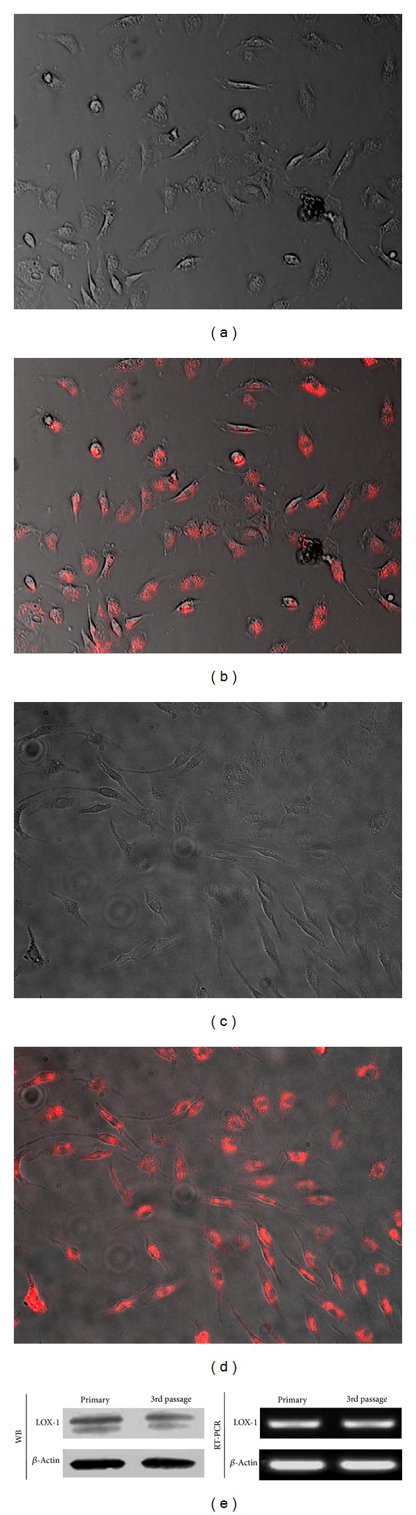
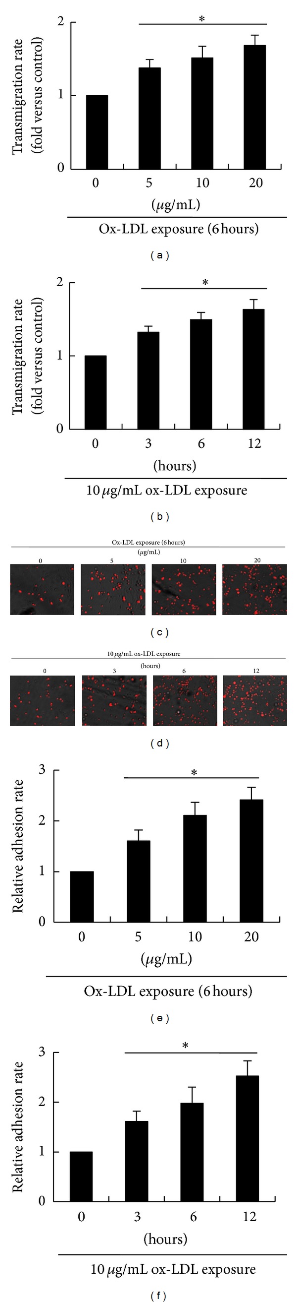
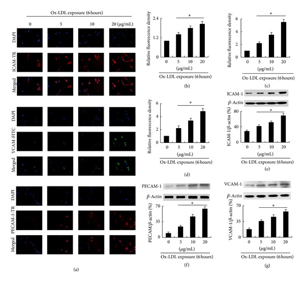
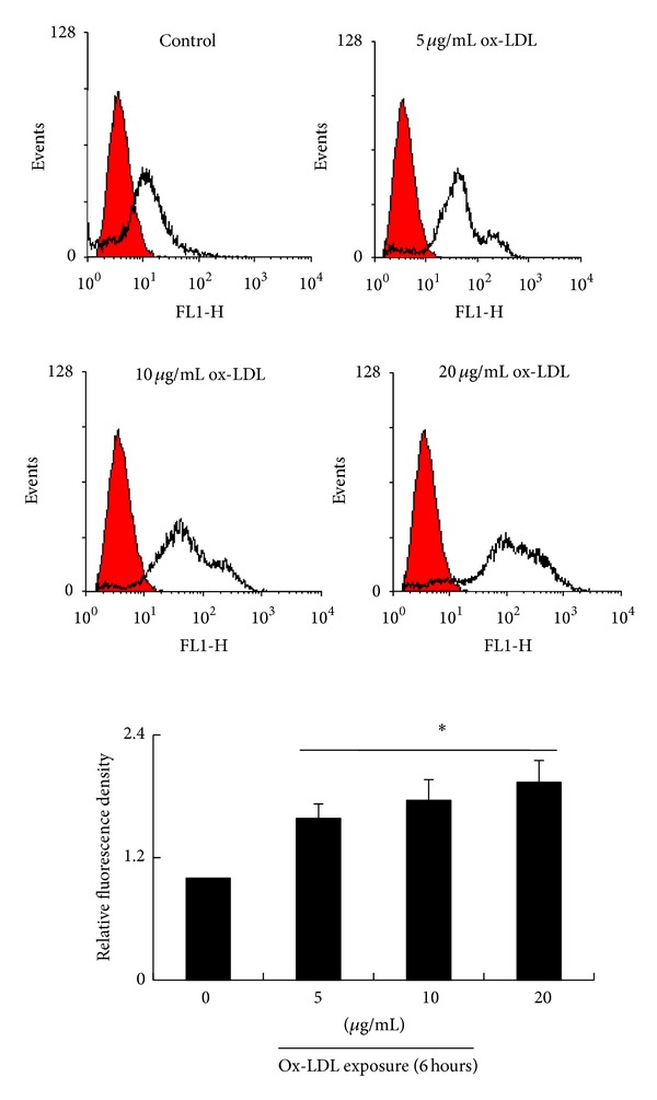
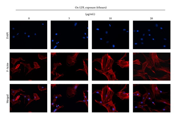
Bone marrow-derived mesenchymal stem cells (bmMSCs) are the most important cell source for stem cell transplant therapy. The migration capacity of MSCs is one of the determinants of the efficiency of MSC-based transplant therapy. Our recent study has shown that low concentrations of oxidized low-density lipoprotein (ox-LDL) can stimulate proliferation of bmMSCs. In this study, we investigated the effects of ox-LDL on bmMSC migration and adhesion, as well as the related mechanisms. Our results show that transmigration rates of bmMSCs and cell-cell adhesion between bmMSCs and monocytes are significantly increased by treatments with ox-LDL in a dose- and time-dependent manner. Expressions of ICAM-1, PECAM-1, and VCAM-1 as well as the levels of intracellular Ca(2+) are also markedly increased by ox-LDL in a dose-dependent manner. Cytoskeleton analysis shows that ox-LDL treatment benefits to spreading of bmMSCs and organization of F-actin fibers after being plated for 6 hours. More interestingly, treatments with ox-LDL also markedly increase expressions of LOX-1, MCP-1, and TGF- β ; however, LOX-1 antibody and MCP-1 shRNA markedly inhibit ox-LDL-induced migration and adhesion of bmMSCs, which suggests that ox-LDL-induced bmMSC migration and adhesion are dependent on LOX-1 activation and MCP-1 expression.
Figures

Similar articles
-
Lectin-like oxidized LDL receptor-1 expresses in mouse bone marrow-derived mesenchymal stem cells and stimulates their proliferation.Exp Cell Res. 2013 Apr 15;319(7):1054-9. doi: 10.1016/j.yexcr.2013.01.021. Epub 2013 Feb 8. Exp Cell Res. 2013. PMID: 23399833
-
Transforming growth factor-beta(1) modulates oxidatively modified LDL-induced expression of adhesion molecules: role of LOX-1.Circ Res. 2001 Dec 7;89(12):1155-60. doi: 10.1161/hh2401.100598. Circ Res. 2001. PMID: 11739280
-
Adhesion molecule expression in fibroblasts: alteration in fibroblast biology after transfection with LOX-1 plasmids.Hypertension. 2005 Sep;46(3):622-7. doi: 10.1161/01.HYP.0000179045.95915.b0. Epub 2005 Aug 22. Hypertension. 2005. PMID: 16116044
-
Oxidized LDL, LOX-1 and atherosclerosis.Cardiovasc Drugs Ther. 2011 Oct;25(5):419-29. doi: 10.1007/s10557-011-6341-5. Cardiovasc Drugs Ther. 2011. PMID: 21947818 Review.
-
Role of Ox-LDL and LOX-1 in Atherogenesis.Curr Med Chem. 2019;26(9):1693-1700. doi: 10.2174/0929867325666180508100950. Curr Med Chem. 2019. PMID: 29737246 Review.
Cited by
-
Macrophage Differentiation from Monocytes Is Influenced by the Lipid Oxidation Degree of Low Density Lipoprotein.Mediators Inflamm. 2015;2015:235797. doi: 10.1155/2015/235797. Epub 2015 Jul 29. Mediators Inflamm. 2015. PMID: 26294848 Free PMC article.
-
Cell membrane damage is involved in the impaired survival of bone marrow stem cells by oxidized low-density lipoprotein.J Cell Mol Med. 2014 Dec;18(12):2445-53. doi: 10.1111/jcmm.12424. Epub 2014 Sep 25. J Cell Mol Med. 2014. PMID: 25256620 Free PMC article.
-
Mesenchymal stromal cells conditioned by peripheral blood mononuclear cells exert enhanced immunomodulation capacities and alleviate a model of Myasthenia Gravis.Stem Cell Res Ther. 2025 Aug 8;16(1):437. doi: 10.1186/s13287-025-04534-9. Stem Cell Res Ther. 2025. PMID: 40781689 Free PMC article.
-
oxLDL promotes podocyte migration by regulating CXCL16, ADAM10 and ACTN4.Mol Med Rep. 2020 Sep;22(3):1976-1984. doi: 10.3892/mmr.2020.11292. Epub 2020 Jun 30. Mol Med Rep. 2020. PMID: 32705248 Free PMC article.
-
CCL13 and human diseases.Front Immunol. 2023 Apr 19;14:1176639. doi: 10.3389/fimmu.2023.1176639. eCollection 2023. Front Immunol. 2023. PMID: 37153575 Free PMC article. Review.
References
-
- Zhang F, Wang C, Jing S, et al. Lectin-like oxidized LDL receptor-1 expresses in mouse bone marrow-derived mesenchymal stem cells and stimulates their proliferation. Experimental Cell Research. 2013;319(7):1054–1059. - PubMed
-
- Lee JK, Jin HK, Endo S, Schuchman EH, Carter JE, Bae J-S. Intracerebral transplantation of bone marrow-derived mesenchymal stem cells reduces amyloid-beta deposition and rescues memory deficits in Alzheimer’s disease mice by modulation of immune responses. Stem Cells. 2010;28(2):329–343. - PubMed
-
- Huang W, Mo X, Qin C, Zheng J, Liang Z, Zhang C. Transplantation of differentiated bone marrow stromal cells promotes motor functional recovery in rats with stroke. Neurological Research. 2013;35(3):320–328. - PubMed
Publication types
MeSH terms
Substances
LinkOut - more resources
Full Text Sources
Other Literature Sources
Miscellaneous
