

Squamous cell carcinoma in exstrophy of the bladder
- PMID: 23956833
- PMCID: PMC3742910
- DOI: 10.4111/kju.2013.54.8.555
Squamous cell carcinoma in exstrophy of the bladder
Abstract
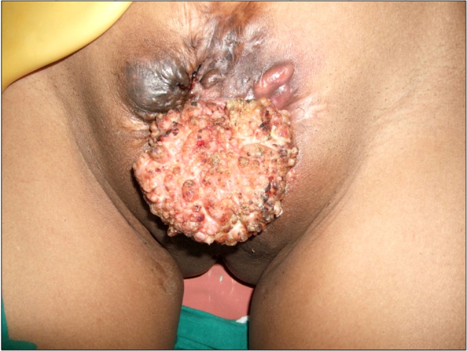
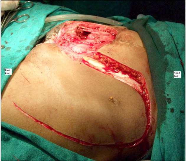
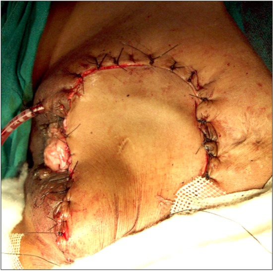
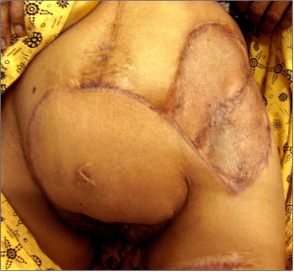
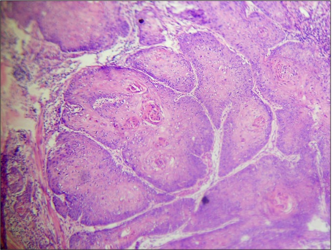
Exstrophy of the bladder is a rare congenital anomaly with an incidence of about 1 per 50,000 newborns. The malignant potential of the exstrophied bladder mucosa is well known; 95% are adenocarcinomas, and 3% to 5% are squamous cell carcinomas. Most of the malignant tumors (60%) associated with an exstrophy of the bladder occur during the fourth and fifth decades of life. Of the remaining, about 20% each occur after 60 years and before 40 years. Here we present a case in which squamous cell carcinoma developed in an unrepaired exstrophy of the bladder. We present the management of the case and a brief review of the literature.
Keywords: Bladder exstrophy; Squamous cell carcinoma; Urinary bladder neoplasms.
Conflict of interest statement
The authors have nothing to disclose.
Figures
Similar articles
-
[Bladder exstrophy with adenocarcinoma in an adult patient: a case report].Pan Afr Med J. 2018 Apr 3;29:197. doi: 10.11604/pamj.2018.29.197.15291. eCollection 2018. Pan Afr Med J. 2018. PMID: 30061975 Free PMC article. French.
-
Oncologic Concerns in An Exstrophied Urinary Bladder - An Indian Scenario.J Clin Diagn Res. 2015 Sep;9(9):XD04-XD05. doi: 10.7860/JCDR/2015/12352.6467. Epub 2015 Sep 1. J Clin Diagn Res. 2015. PMID: 26500998 Free PMC article.
-
Anterior pelvic exenteration for exstrophic bladder adenocarcinoma: Case report and review.Int J Surg Case Rep. 2016;25:13-5. doi: 10.1016/j.ijscr.2016.05.022. Epub 2016 Jun 1. Int J Surg Case Rep. 2016. PMID: 27288750 Free PMC article.
-
Embryology and bony and pelvic floor anatomy in the bladder exstrophy-epispadias complex.Semin Pediatr Surg. 2011 May;20(2):66-70. doi: 10.1053/j.sempedsurg.2010.12.011. Semin Pediatr Surg. 2011. PMID: 21453848 Review.
-
Female bladder exstrophy: report of 2 unique cases and review of the literature.Postgrad Med. 2012 May;124(3):37-41. doi: 10.3810/pgm.2012.05.2546. Postgrad Med. 2012. PMID: 22691897 Review.
Cited by
-
Pesticides and Bladder Cancer: Mechanisms Leading to Anti-Cancer Drug Chemoresistance and New Chemosensitization Strategies.Int J Mol Sci. 2023 Jul 13;24(14):11395. doi: 10.3390/ijms241411395. Int J Mol Sci. 2023. PMID: 37511154 Free PMC article. Review.
-
Primary urothelial carcinoma of an ileal conduit; six decades after childhood bladder exstrophy surgery: a rare and late complication.World J Surg Oncol. 2025 May 31;23(1):211. doi: 10.1186/s12957-025-03798-y. World J Surg Oncol. 2025. PMID: 40450330 Free PMC article.
-
Squamous cell carcinoma at bladder exstrophy site after early cystectomy.Pediatr Surg Int. 2015 Nov;31(11):1107-10. doi: 10.1007/s00383-015-3725-9. Epub 2015 Jul 14. Pediatr Surg Int. 2015. PMID: 26169528
-
[Bladder exstrophy with adenocarcinoma in an adult patient: a case report].Pan Afr Med J. 2018 Apr 3;29:197. doi: 10.11604/pamj.2018.29.197.15291. eCollection 2018. Pan Afr Med J. 2018. PMID: 30061975 Free PMC article. French.
-
Adenocarcinoma mucinosum of extrophy bladder: A rare case report.Int J Surg Case Rep. 2021 Nov;88:106493. doi: 10.1016/j.ijscr.2021.106493. Epub 2021 Oct 14. Int J Surg Case Rep. 2021. PMID: 34717273 Free PMC article.
References
-
- Lattimer JK, Smith MJ. Exstrophy closure: a followup on 70 cases. J Urol. 1966;95:356–359. - PubMed
-
- de Riese W, Warmbold H. Adenocarcinoma in extrophy of the bladder: a case report and review of the literature. Int Urol Nephrol. 1986;18:159–162. - PubMed
-
- Gupta S, Gupta IM. Ectopia vesicae complicated by squamous cell carcinoma. Br J Urol. 1976;48:244. - PubMed
-
- Facchini V, Gadducci A, Colombi L, Ceccarelli P, Lamacchia M, Simi U, et al. Carcinoma developing in bladder exstrophy. Case report. Br J Obstet Gynaecol. 1987;94:795–797. - PubMed
-
- Serretta V, Pomara G, Piazza F, Gange E. Pure squamous cell carcinoma of the bladder in western countries: report on 19 consecutive cases. Eur Urol. 2000;37:85–89. - PubMed
LinkOut - more resources
Full Text Sources
Other Literature Sources
