

Amyloid fiber formation in human γD-Crystallin induced by UV-B photodamage
- PMID: 23957864
- PMCID: PMC3859806
- DOI: 10.1021/bi4008353
Amyloid fiber formation in human γD-Crystallin induced by UV-B photodamage
Abstract
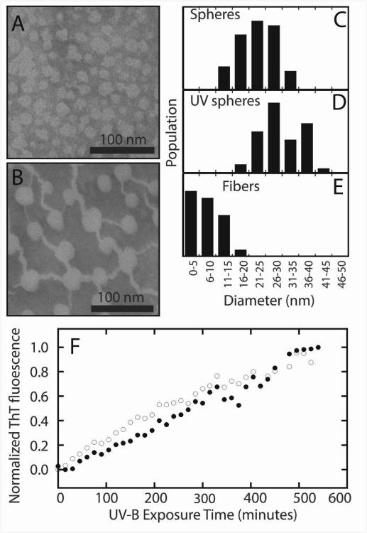
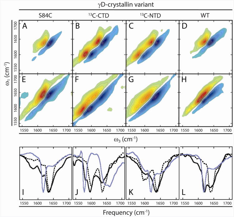
γD-Crystallin is an abundant structural protein of the lens that is found in native and modified forms in cataractous aggregates. We establish that UV-B irradiation of γD-Crystallin leads to structurally specific modifications and precipitation via two mechanisms: amorphous aggregates and amyloid fibers. UV-B radiation causes cleavage of the backbone, in large measure near the interdomain interface, where side chain oxidations are also concentrated. 2D IR spectroscopy and expressed protein ligation localize fiber formation exclusively to the C-terminal domain of γD-Crystallin. The native β-sandwich domains are not retained upon precipitation by either mechanism. The similarities between the amyloid forming pathways when induced by either UV-B radiation or low pH suggest that the propensity for the C-terminal β-sandwich domain to form amyloid β-sheets determines the misfolding pathway independent of the mechanism of denaturation.
Figures
References
-
- Wang Y, King JA. Protein Misfolding Diseases. John Wiley & Sons, Inc; 2010. Cataract as a protein-aggregation disease; pp. 487–515.
-
- Bloemendal H, de Jong W, Jaenicke R, Lubsen NH, Slingsby C, Tardieu A. Ageing and vision: structure, stability and function of lens crystallins. Prog Biophys Mol Biol. 2004;86:407–485. - PubMed
-
- Truscott RJW. Age-related nuclear cataract—oxidation is the key. Experimental Eye Research. 2005;80:709–725. - PubMed
-
- Hanson SRA, Hasan A, Smith DL, Smith JB. The Major in vivo Modifications of the Human Water-insoluble Lens Crystallins are Disulfide Bonds, Deamidation, Methionine Oxidation and Backbone Cleavage. Experimental Eye Research. 2000;71:195–207. - PubMed
Publication types
MeSH terms
Substances
Grants and funding
LinkOut - more resources
Full Text Sources
Other Literature Sources
