Review
. 2013 Sep;34(9):423-30.
doi: 10.1016/j.it.2013.07.001. Epub 2013 Aug 16.

Impact of the gut microbiome on mucosal inflammation

Affiliations
Review

Impact of the gut microbiome on mucosal inflammation

Warren Strober. Trends Immunol. 2013 Sep.

Abstract

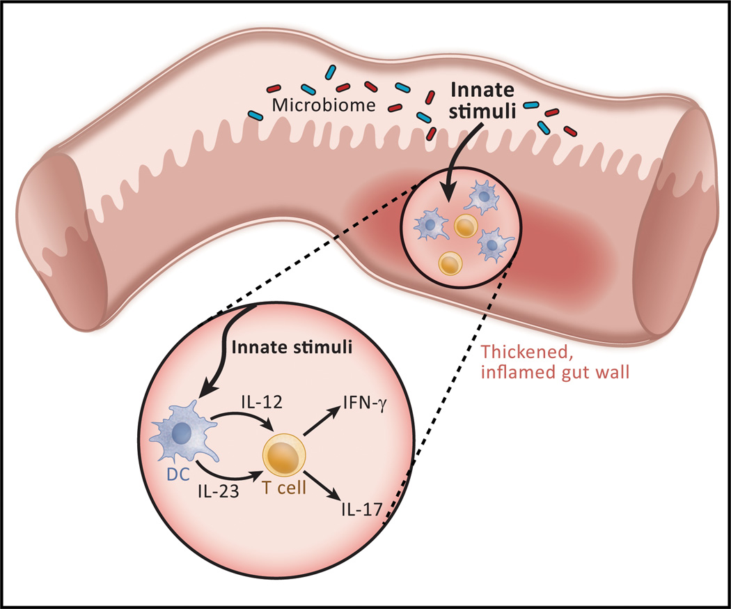
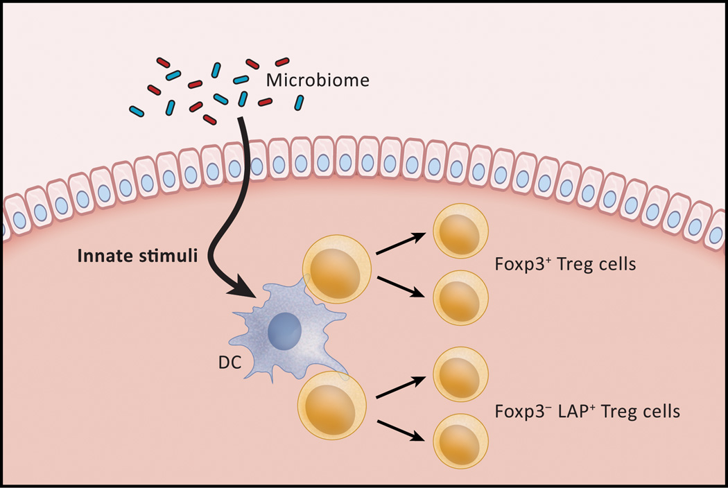
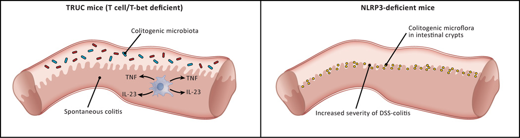
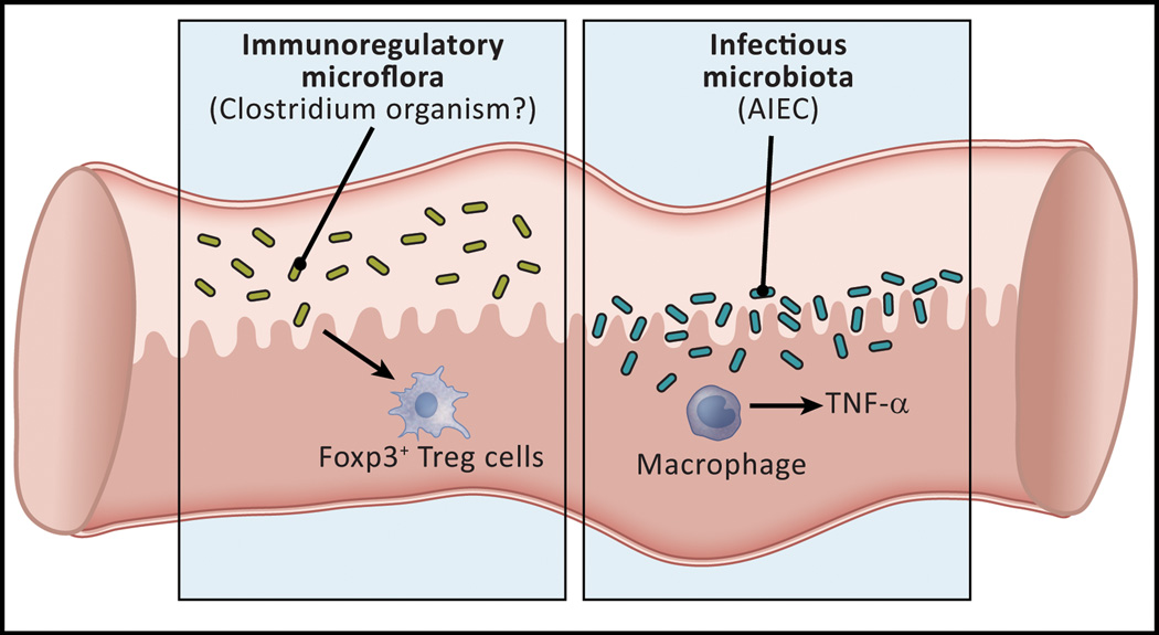
In the past 10 years it has become increasingly apparent that the gut microbiome has profound effects on the immune system to which it is juxtaposed, the mucosal immune system. Here, I explore recent studies in which the effects of the microbiota expand or facilitate anti-inflammatory or regulatory immunological machinery or which favor development of proinflammatory immunological machinery in this system. I then focus on how these opposing processes play out in inflammatory bowel disease (IBD); a disease in which normal immune homeostasis is disturbed and inflammation takes hold.

Keywords: Anti-inflammatory organisms; Gut microbiome; Inflammatory Bowel Disease; Mucosal regulatory T cells; Proinflammatory (colitogenic organisms).


Figures

Figure 1
Figure 1
The basis of Crohn’s disease, a major form of inflammatory bowel disease. The consensus view of Crohn’s disease pathogenesis is that the latter consists of, at least in part, an excessive immunologic response to some component of the microbiota existing in the bowel lumen, most likely a MAMP (microbial-associated molecular pattern) interacting with either a TLR or an NLR. Thus, a MAMP stimulation gives rise to production of cytokines such as IL-12 or IL-23 that induce T cell differentiation into Th1 or Th17 cells, the ultimate effectors of the inflammation. The excessive response could be due to a direct disturbance in the induction of effector cells or to an indirect disturbance in the regulatory cells that control such induction. The mechanism underlying ulcerative colitis is thought to be similar although the cellular processes resulting from the excessive response is different.
Figure 2
Figure 2
Homeostatic regulation of mucosal responses. Although the epithelial cell layer of the intestine is a barrier to the entry of luminal materials, it is porous enough to allow entry of microbiome-derived components that mediate the development of regulatory T cells. As shown in this Figure, innate stimuli (TLR or NLR ligands) can interact with dendritic cells in the lamina propria and these, in turn, activate regulatory cells that maintain a quiescent state in the unperturbed intestine despite the presence of adjacent microbiota. In addition, innate stimuli may interact directly with the regulatory cell as well. Two types of regulatory cells have been described: a Tr1-like (IL-10-dependent) cell that bears surface TGF-β and an (IL-10-independent) Foxp3+ cell. Under conditions in which the epithelial barrier is made more porous these cells increase in number, but in this case their function is overwhelmed by a strong pro-inflammatory stimulus.
Figure 3
Figure 3
Two types of “colitogenic” microbiota. Studies of murine models of GI inflammation have identified two types of “colitogenic” microbiota, i.e., microbiota that cause colitis in a normal host. Truly colitogenic microbiota, so far only identified in TRUC mice (see text) are organisms that cause spontaneous colitis in co-housed normal mice. Partially, colitogenic microbiota are represented by Prevotella organisms that proliferate in the intestinal crypts of mice lacking epithelial cells that produce NLRP3 and therefore are deficient in IL-18 production. These organisms are partially colitogenic because they cause intensification of a pre-existing colitis and not de novo colitis
Figure 4
Figure 4
The gut microflora may cause or aggravate Crohn’s disease by defective induction of regulatory T cells or by infection of the mucosa and the induction of inflammatory cytokines. Studies of mice and humans with GI inflammation have led to the identification of two kinds of microflora that may cause or aggravate the inflammation of Crohn’s disease. In the first category are organisms in the Firmacute phylum represented by Faecalibacterium prausnitzii, a clostridial organism that may be related to those shown in murine systems to induce regulatory T cells in the gut. In the second category is an E.coli organism known as “adherent-invasive” E. coli (AIEC) that lacks pathogenic properties in normals but that colonizes the small intestine of some patients with Crohn’s disease. This organism invades macrophages and induces the latter cells to produce pro-inflammatory cytokines.

Similar articles

Cited by

References

    1. Strober W, Fuss IJ, Blumberg RS. The immunology of mucosal models of inflammation. Annu. Rev. Immunol. 2001;20:495–549. - PubMed
    1. Kaser A, Zeissig S, Blumberg RS. Inflammatory bowel disease. Annu. Rev. Immunol. 2010;28:573–621. - PMC - PubMed
    1. Strober W, Fuss I, Mannon P. The fundamental basis of inflammatory bowel disease. J. Clin. Invest. 2007;117:514–521. - PMC - PubMed
    1. Strober W, Watanabe T. NOD2, an intracellular innate immune sensor involved in host defense and Crohn’s disease. Mucosal Immunol. 2011;4:484–495. - PMC - PubMed
    1. Saitoh T, Fujita N, Jang MH, Uematsu S, Yang BG, Satoh T, Omori H, Noda T, Yamamoto N, Komatsu M, Tanaka K, Kawai T, Tsujmura T, Takeuchi O, Yoshimori T, Akira S. Loss of the autophagy protein Atg16L1 enhances endotoxin-induced IL-1beta production. Nature. 2008;456:264–268. - PubMed