Review
. 2013 Dec;23(12):620-33.
doi: 10.1016/j.tcb.2013.07.006. Epub 2013 Aug 16.

Evading apoptosis in cancer

Affiliations
Review

Evading apoptosis in cancer

Kaleigh Fernald et al. Trends Cell Biol. 2013 Dec.

Abstract

Carcinogenesis is a mechanistically complex and variable process with a plethora of underlying genetic causes. Cancer development comprises a multitude of steps that occur progressively starting with initial driver mutations leading to tumorigenesis and, ultimately, metastasis. During these transitions, cancer cells accumulate a series of genetic alterations that confer on the cells an unwarranted survival and proliferative advantage. During the course of development, however, cancer cells also encounter a physiologically ubiquitous cellular program that aims to eliminate damaged or abnormal cells: apoptosis. Thus, it is essential that cancer cells acquire instruments to circumvent programmed cell death. Here we discuss emerging evidence indicating how cancer cells adopt various strategies to override apoptosis, including amplifying the antiapoptotic machinery, downregulating the proapoptotic program, or both.

Keywords: BH3; MOMP; caspase; phosphorylation; ubiquitination.


Figures

Figure 1
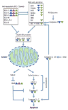
Figure 1. Apoptosis is triggered in response to internal or external stimuli
The intrinsic pathway to apoptosis is initiated by activation of BH3-only protein. Among the BH3-only proteins, whose expression is often transcriptionally induced following an apoptotic stimulus, BID is unique in that it is activated by caspase cleavage (by caspases-2 and -8/10); cleaved BID, termed tBID. BH3 proteins trigger the activation of multi-BH domain proteins by direct binding or by inhibition of anti-apoptotic BCL-2 (B-cell lymphoma 2) family proteins. Once activated, multi-BH domain proteins form oligomers that induce MOMP. MOMP prompts pro-apoptotic proteins SMAC and cytochrome c to be released to the cytoplasm, where cytochrome c induces the formation of the Apoptosome. The initiator caspase-9 activated within the Apoptosome initiates the activation of executioner caspase-3 and -7, resulting in apoptosis. Of note, caspase-2 is activated via formation of the PIDDosome, a complex which consists of the adaptor proteins PIDD (p53-induced death domain protein) and RAIDD (RIP-associated protein with a death domain). However, despite identification of this complex, the precise mechanism of caspase-2 activation remains elusive. In addition, only a few cellular substrates of caspase-2, including BID, have been identified. Colored triangles denote that the indicated gene or gene product may be transcriptionally (blue), translationally (red), and post-translationally (green) up- or down-regulated in cancer cells (see text and Table 1).
Figure 2
Figure 2. Oncogenic kinase signaling regulates MCL-1, BIM, and PUMA
Oncogenic kinases regulate BIM, PUMA, and MCL-1 by changing gene expression, protein stability, and protein functions by activating two major pro-survival pathways: the AKT and ERK (Extracellular signal-regulated kinases) pathways. Pro-survival and pro-apoptotic pathways are denoted in blue and red, respectively.
Figure 3
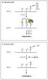
Figure 3. MCL-1 protein stability is controlled by multiple kinases
(A) MCL-1 can be phosphorylated at T163 by several kinases including GSK3. AKT inhibits GSK3. Thus, in low levels of AKT, phosphorylation by GSK3 sequentially occurs at S155/S159, targeting MCL-1 for degradation by E3 ligases, SCFβ-TrCP, SCFFBW7 (F-box/WD repeat-containing protein 7), and TRIM17. (B) In cancer cells with high levels of AKT and ERK, ERK is likely to be the main kinase that phosphorylates MCL-1. Phosphorylation does not continue to S155/S159 and MCL-1 is stabilized.

References

    1. Martinou JC, Youle RJ. Mitochondria in apoptosis: Bcl-2 family members and mitochondrial dynamics. Dev Cell. 2011;21:92–101. - PMC - PubMed
    1. Strasser A, et al. Deciphering the rules of programmed cell death to improve therapy of cancer and other diseases. EMJO J. 2011;30:3667–3683. - PMC - PubMed
    1. Rautureau GJ, et al. The restricted binding repertoire of Bcl-B leaves Bim as the universal BH3-only prosurvival Bcl-2 protein antagonist. Cell Death Dis. 2012;13:e443. - PMC - PubMed
    1. Hübner A, et al. Multisite phosphorylation regulates Bim stability and apoptotic activity. Mol Cell. 2008;30:415–425. - PMC - PubMed
    1. Bean GR, et al. PUMA and BIM Are Required for Oncogene Inactivation-Induced Apoptosis. Sci Sinal. 2013:6,ra20. - PMC - PubMed

Publication types

Substances