. 2013 Sep 10:186:92-103.
doi: 10.1016/j.regpep.2013.08.001. Epub 2013 Aug 17.

Keratinocytes express cytokines and nerve growth factor in response to neuropeptide activation of the ERK1/2 and JNK MAPK transcription pathways

Affiliations

Keratinocytes express cytokines and nerve growth factor in response to neuropeptide activation of the ERK1/2 and JNK MAPK transcription pathways

Xiaoyou Shi et al. Regul Pept. .

Abstract

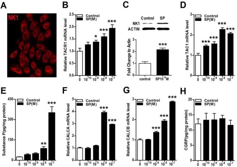
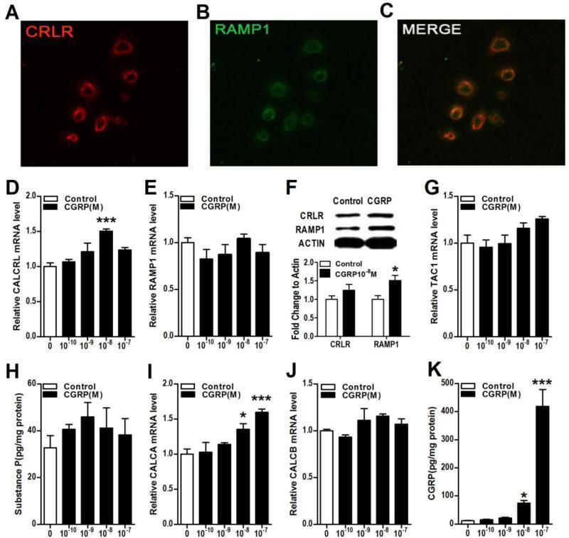
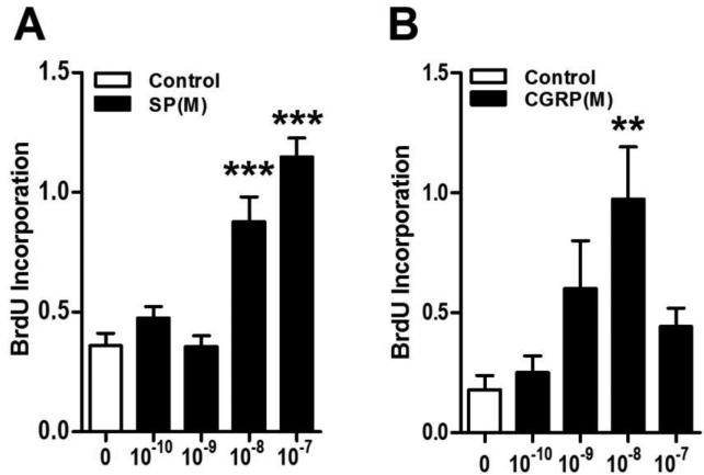
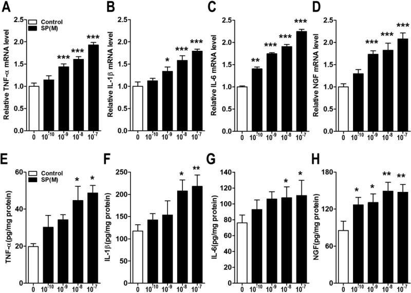
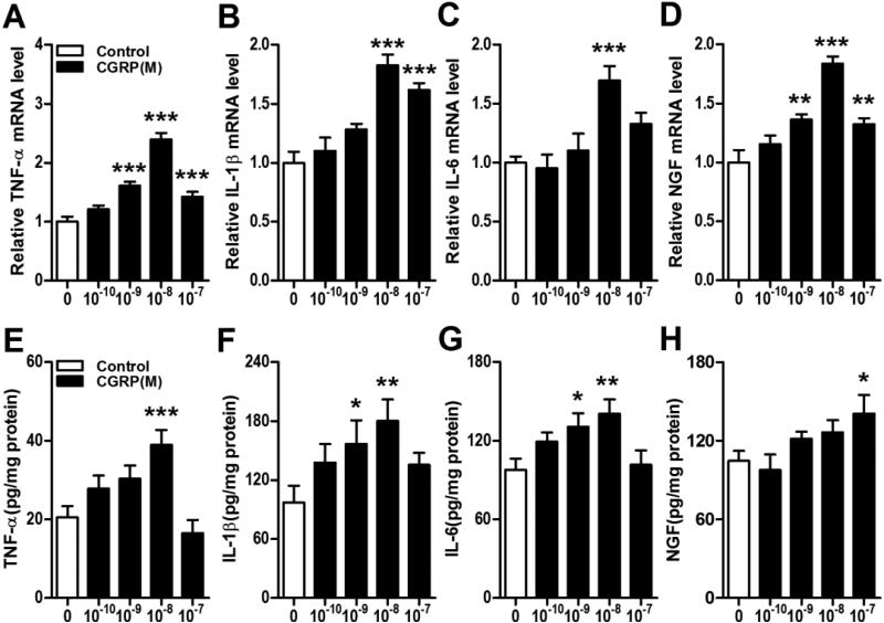
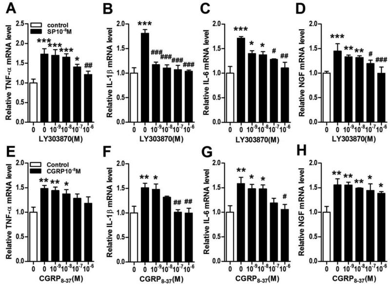
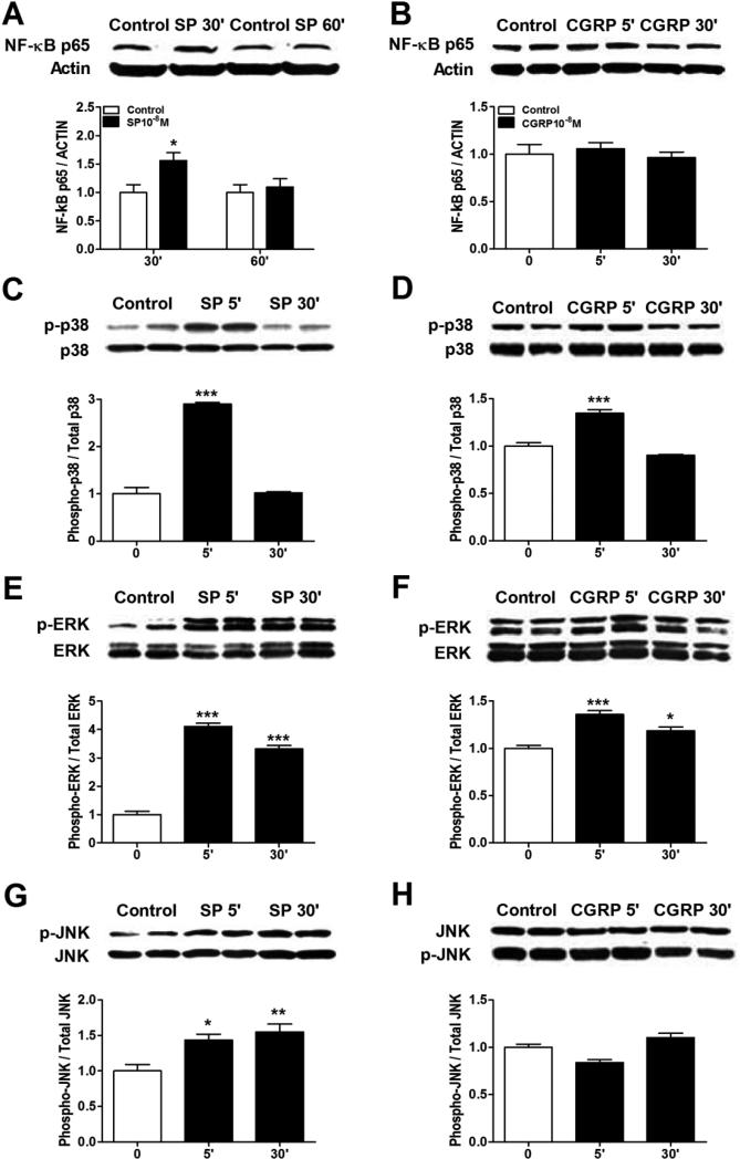
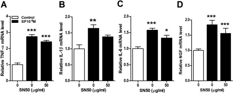
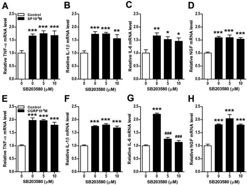
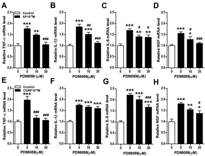
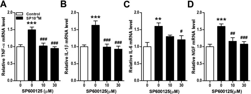
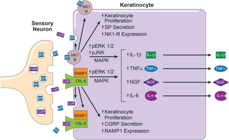
Sensory neurons innervating the skin can release neuropeptides that are believed to modulate cellular proliferation, wound healing, pigmentation, and keratinocyte innate immune responses. While the ability of neuropeptides to stimulate keratinocyte production of inflammatory mediators has been demonstrated, there is no information concerning the mechanisms by which neuropeptide activation of keratinocyte cell surface receptors ultimately leads to the up-regulation of mediator production. In this study we used a keratinocyte cell line to identify the presence of substance P (SP) and calcitonin gene-related peptide (CGRP) receptors on keratinocytes and examined the effects of SP and CGRP stimulation on keratinocyte neuropeptide signaling, cell proliferation, and interleukin-1β (IL-1β), interleukin-6 (IL-6), tumor necrosis factor α (TNF-α), and nerve growth factor (NGF) expression. Neuropeptide stimulation caused an up-regulation of neuropeptide receptor expression in keratinocytes and a dramatic increase in keratinocyte secretion of SP and CGRP, suggesting possible autocrine or paracrine stimulatory effects and amplification of neuropeptide signaling. Both SP and CGRP concentration-dependently stimulated cellular proliferation and the expression and secretion of inflammatory cytokines and NGF in keratinocytes. SP also activated all 3 families of mitogen activated protein kinase (MAPK) and nuclear factor κB (NFκB) in keratinocytes, while CGRP only activated p38 and extracellular signal related kinase1/2 (ERK1/2) MAPKs. Neuropeptide stimulated inflammatory mediatory production in keratinocytes was reversed by ERK1/2 and JNK inhibitors. The current study is the first to observe; 1) that CGRP stimulates keratinocyte expression of CGRP and its receptor complex, 2) that SP and CGRP stimulate IL-6 and TNF-α secretion in keratinocytes, 3) that SP activated all three MAPK families and the NFκB transcriptional signaling pathway in keratinocytes, and 4) that SP and CGRP stimulated inflammatory mediator production in keratinocytes is dependent on ERK1/2 and JNK activation. These studies provide evidence suggesting that disruption of ERK1/2 and JNK signaling may potentially be an effective therapy for inflammatory skin diseases and pain syndromes mediated by exaggerated sensory neuron-keratinocyte signaling.

Keywords: Calcitonin gene-related peptide; Cytokines; Keratinocyte; Mitogen activated protein kinases; Nerve growth factor; Substance P.


Figures

Figure 1
Figure 1. Substance P (SP) treatment increased keratinocyte NK1 receptor expression and secretion of SP
The substance P (SP) receptor NK1 was expressed in keratinocytes in vitro, and this expression was up-regulated after SP treatment. SP treatment also increased the gene and protein expression of SP and the gene expression of calcitonin gene-related peptide (CGRP) keratinocytes. Immunostaining demonstrated NK1 protein (red fluorescence) present in the cytoplasm and on the membrane of keratinocytes (A). SP concentration-dependently increased expression of its NK1 receptor gene (TACR1) in keratinocytes at 3 h after treatment, as measured by real time PCR (B). Western blot demonstrates that SP (10−8M) treatment increased NK1 protein levels in keratinocytes at 24 h after treatment (C). SP treatment also concentration-dependently increased the expression of the SP TAC1 gene in keratinocytes at 3h, as measured by real time PCR (D). When keratinocytes were treated with various concentrations of SP for 3h, then washed 3 times in fresh medium, and then incubated in fresh medium for 21h, there was a 15-fold increase in medium SP levels, as measured by EIA (E). SP treatment also increased expression of the CGRP genes CALCA (F) and CALCB (G) in keratinocytes at 3 h. When keratinocytes were treated with various concentrations of SP for 3h, then washed 3 times in medium, and then incubated in fresh medium for 21h, there was no increase in medium levels of CGRP, as measured by EIA (H). Values are means ± SE. * P<0.05, ** P<0.01, and *** P<0.001 vs vehicle treated control cells. All experiments were repeated 3-4 times.
Figure 2
Figure 2. CGRP treatment increased keratinocyte RAMP1 expression and secretion of CGRP
The CGRP receptor components calcitonin receptor-like receptor (CRLR) and receptor activity-modifying protein 1 (RAMP1) are expressed in keratinocytes in vitro and this expression is up-regulated by CGRP treatment. CGRP treatment had no effect on SP gene or protein levels but did stimulate CGRP gene expression and protein secretion in keratinocytes. Immunostaining demonstrated CRLR protein (red) and RAMP1 protein (green) co-expressing in the cytoplasm and on the membrane of keratinocytes (A-C). CGRP treatment increased expression of its CRLR receptor gene (CALCRL) but not its RAMP1 receptor gene (RAMP1) at 3 h after treatment, as measured by real time PCR (D, E). Western blot demonstrates that CGRP (10−8M) increased RAMP1 protein level in keratinocytes at 24 h after treatment (F). CGRP treatment did not change the expression of the SP TAC1 gene at 3h, as measured by real time PCR (G). When keratinocytes were treated with various concentrations of CGRP for 3h, then washed 3 times in fresh medium, and then incubated in fresh medium for 21h, there was no increase in medium SP levels, as measured by EIA (H). CGRP treatment increased the expression of the CGRP gene CALCA (I), but not CALCB (J) at 3 h. When keratinocytes were treated with various concentrations of CGRP for 3h, then washed 3 times in fresh medium, and then incubated in fresh medium for 21h, there was a 44-fold increase in medium CGRP levels, as measured by EIA (K). Values are means ± SE. * P<0.05, ** P<0.01, and *** P<0.001 vs vehicle treated control cells. All experiments were repeated 3-4 times.
Figure 3
Figure 3. SP and CGRP stimulated keratinocyte cellular proliferation
SP (A) and CGRP (B) concentration-dependently stimulated cell proliferation in keratinocytes in vitro, as measured by BrdU incorporation over a 24 hour period. Values are means ± SE. * P<0.05, ** P<0.01 and *** P<0.001 and vs vehicle treated control cells. These experiments were repeated 3 times.
Figure 4
Figure 4. SP treatment stimulated keratinocyte expression of inflammatory mediators
SP concentration-dependently up-regulated tumor necrosis factor α (TNF-α), interleukin 1 (IL-1), interleukin 6 (IL-6), and nerve growth factor (NGF) gene expression in keratinocytes at 3 h, as measured by real time PCR (A-D) and similarly increased cytokine and NGF protein secretion into the media over 24 hours after SP treatment, as measured by EIA (E-H). Values are means ± SE. * P<0.05, ** P<0.01, and *** P<0.001 vs vehicle treated control cells. These experiments were repeated 3-4 times.
Figure 5
Figure 5. CGRP stimulated keratinocyte expression of inflammatory mediators
CGRP concentration-dependently up-regulated TNF-α, IL-1, IL-6, and NGF gene expression in keratinocytes at 3 h, as measured by real time PCR (A-D) and similarly increased cytokine and NGF protein secretion into the media over 24 hours after SP treatment, by EIA (E-H). Values are means ± SE. * P<0.05, ** P<0.01, and *** P<0.001 vs vehicle treated control cells. These experiments were repeated 3-4 times.
Figure 6
Figure 6. Inflammatory mediator expression in keratinocytes required neuropeptide receptor activation
SP (10−8M) induced up-regulation of cytokine and NGF gene expression in keratinocytes was concentration-dependently inhibited by the SP NK1 receptor antagonist LY303870, as measured by real time PCR at 3 h after SP treatment (A-D). Similarly, CGRP (10−8M) evoked up-regulation of cytokine gene expression in keratinocytes was concentration-dependently inhibited by the CGRP receptor antagonist CGRP8-37, as measured by real time PCR at 3 h after CGRP treatment (E-G). CGRP8-37 treatment had no significant effect on CGRP stimulated NGF gene expression in keratinocytes (H). Values are means ± SE. * P<0.05, ** P<0.01, and *** P<0.001 vs vehicle treated control cells. #P<0.05, ## P<0.01 and ### P<0.001 vs SP or CGRP treated cells. These experiments were repeated 3-4 times.
Figure 7
Figure 7. Neuropeptides induced NF-κB and MAPK activation in keratinocytes
Western blot was used to evaluate the stimulatory effects of SP and CGRP treatment on various transcriptional pathways in keratinocytes. SP (10−8M) induced NF-κB p65 nuclear translocation in keratinocytes at 30 min after treatment (A), but CGRP (10−8M) had no effect on NF-κB nuclear translocation (B). Both SP (C) and CGRP (D) caused the rapid phosphorylation of p38 mitogen activated protein kinase (MAPK) at 5 min, but CGRP had a less robust effect. Similarly, Both SP (E) and CGRP (F) caused the rapid phosphorylation of extracellular signal related kinases 1/2 (ERK1/2) MAPK at 5 and 30 min, but again CGRP had a less robust effect. SP (G) treatment induced moderate phosphorylation of c-Jun N-terminal kinases (JNK) MAPK at 5 and 30 min after treatment, but CGRP (H) had no effect. These experiments were repeated 3-4 times.
Figure 8
Figure 8. NF-κB activation was not required for SP stimulation of keratinocyte inflammatory mediator expression
The NF-kB inhibitor SN50 (50 ug/ml) did not inhibit SP (10−8M) stimulated TNF (A), IL-1 (B), IL-6 (C), and NGF (D) gene expression in keratinocytes at 3 h, measured by real time PCR. The same concentration of SN50 (50 ug/ml) that was ineffective in blocking SP stimulation of cytokine and NGF expression completely blocked SP stimulated NF-κB p65 nuclear translocation (data not shown), suggesting that other transcription pathways are responsible for SP induced inflammatory mediator expression. There were no significant differences between the SP and SP + SN50 groups (black bars) for any inflammatory mediator. CGRP treatment did not induce NF-κB nuclear translocation (Fig. 7), thus CGRP was not tested against SN50. Values are means ± SE. * P<0.05, ** P<0.01, and *** P<0.001 vs vehicle treated control cells.
Figure 9
Figure 9. Neuropeptide stimulation of keratinocyte inflammatory mediator expression does not require p38 MAPK activation
The p38 MAPK inhibitor SB203580 was used to demonstrate that SP and CGRP induced cytokine and NGF expression in keratinocytes does not require p38 activation. SB203580 had no effect on SP (10−8M, black bars) stimulation of TNF (A), IL-1 (B), IL-6 (C), and NGF (D) gene expression in keratinocytes at 3 h, measured by real time PCR. Similarly, SB203580 had no effect on CGRP (10−8M, black bars) stimulation of TNF (E), IL-1 (F), and NGF (H) gene expression in keratinocytes at 3 h, measured by real time PCR. SB203580 did inhibit CGRP stimulation of IL-6 (G) expression, suggesting that the p38 kinase activation may contribute to CGRP stimulation of IL-6 expression in keratinocytes. Values are means ± SE. * P<0.05, ** P<0.01, and *** P<0.001vs vehicle treated control cells. # P<0.05, ## P<0.01, and ### P<0.001 vs SP or CGRP treated cells. These experiments were repeated 3-4 times.
Figure 10
Figure 10. Neuropeptide stimulation of keratinocyte inflammatory mediator expression requires ERK MAPK activation
The ERK1/2 MAPK inhibitor PD98059 was used to demonstrate that SP and CGRP induced cytokine and NGF expression in keratinocytes required ERK1/2 activation. PD98059 concentration-dependently inhibited SP (10−8M, black bars) stimulation of TNF (A), IL-1 (B), IL-6 (C), and NGF (D) gene expression in keratinocytes at 3 h, measured by real time PCR. Similarly, PD98059 concentration-dependently inhibited CGRP (10−8M, black bars) stimulation of TNF (E), IL-6 (G), and NGF (H) gene expression in keratinocytes at 3 h, measured by real time PCR. There was no PD98059 inhibition of CGRP stimulation of IL-1 (F) expression, suggesting that other protein kinases or signaling pathways may mediate CGRP stimulation of IL-1 expression in keratinocytes. Values are means ±SE. * P<0.05, ** P<0.01, and *** P<0.001 vs vehicle treated control cells. # P<0.05, ## P<0.01, and ### P<0.001 vs SP or CGRP treated cells. These experiments were repeated 3-4 times.
Figure 11
Figure 11. SP stimulation of keratinocyte inflammatory mediator expression requires ERK MAPK activation
The JNK MAPK inhibitor SP600125 was used to demonstrate that SP induced cytokine and NGF expression in keratinocytes required JNK activation. SP600125 completely blocked SP (10−8M, black bars) stimulation of TNF (A), IL-1 (B), IL-6 (C), and NGF (D) gene expression in keratinocytes at 3 h, measured by real time PCR. CGRP treatment did not induce JNK activation (Fig. 7), thus CGRP was not tested against a SP600125. Values are means ± SE. * P<0.05, ** P<0.01, and *** P<0.001 vs vehicle treated control cells. # P<0.05, ## P<0.01, and ### P<0.001 vs SP treated cells. These experiments were repeated 3-4 times.
Figure 12
Figure 12. This schematic summarizes the results of these experiments
Cutaneous primary sensory afferents release SP and CGRP in the epidermis. These neuropeptides then diffuse through the interstitial space to bind and activate their cognate receptors on the keratinocyte cell surface, the SP NK1 receptor and the CGRP receptor dimer complex of CRLR and RAMP1. Keratinocyte NK1 receptor activation stimulates cellular proliferation, SP expression and secretion, NK1 receptor expression, and the phosphorylation and activation of ERK 1/2 and JNK MAPK intracellular transcription factors that stimulate TNFα, IL-1β, IL-6, and NGF expression and secretion. Similarly, activation of the keratinocyte CGRP receptor dimer complex stimulates keratinocyte proliferation, CGRP expression and secretion, RAMP1 receptor expression, and the phosphorylation and activation of ERK 1/2 MAPK, an intracellular transcription factor that up-regulates TNFα, IL-6, and NGF expression and secretion. Keratinocyte secreted inflammatory cytokines and NGF can directly activate their cognate receptors expressed on cutaneous sensory afferent neurons, with subsequent pain sensitization. Keratinocyte secreted inflammatory mediators also can activate various cellular components of the innate and adaptive immune systems, thus supporting the development of inflammatory skin diseases and nociceptive sensitization. CGRP, calcitonin gene-related peptide; SP, substance P; NK1-R, neurokinin 1 receptor; CRL-R, calcitonin receptor –like receptor; RAMP1 receptor activity-modifying protein; MAPK, mitogen activated protein kinases; ERK1/2; extracellular signal related kinases 1/2; JNK, c-Jun N-terminal kinases; IL, interleukin; TNF α, tumor necrosis factor α; NGF, nerve growth factor.

References

    1. da Silva L, Carvalho E, Cruz MT. Role of neuropeptides in skin inflammation and its involvement in diabetic wound healing. Expert Opin Biol Ther. 2010;10:1427–39. - PubMed
    1. Roosterman D, Goerge T, Schneider SW, Bunnett NW, Steinhoff M. Neuronal control of skin function: the skin as a neuroimmunoendocrine organ. Physiol Rev. 2006;86:1309–79. - PubMed
    1. Dallos A, Kiss M, Polyanka H, Dobozy A, Kemeny L, Husz S. Effects of the neuropeptides substance P, calcitonin gene-related peptide, vasoactive intestinal polypeptide and galanin on the production of nerve growth factor and inflammatory cytokines in cultured human keratinocytes. Neuropeptides. 2006;40:251–63. - PubMed
    1. Burbach GJ, Kim KH, Zivony AS, Kim A, Aranda J, Wright S, Naik SM, Caughman SW, Ansel JC, Armstrong CA. The neurosensory tachykinins substance P and neurokinin A directly induce keratinocyte nerve growth factor. J Invest Dermatol. 2001;117:1075–82. - PubMed
    1. Shi X, Wang L, Li X, Sahbaie P, Kingery WS, Clark JD. Neuropeptides contribute to peripheral nociceptive sensitization by regulating interleukin-1beta production in keratinocytes. Anesth Analg. 2011;113:175–83. - PMC - PubMed

Publication types

MeSH terms

Substances