. 2013 Dec;89(6):1103-16.
doi: 10.4269/ajtmh.13-0205. Epub 2013 Aug 19.

Characterization of three novel adhesins of Leptospira interrogans

Affiliations

Characterization of three novel adhesins of Leptospira interrogans

Gabriela H Siqueira et al. Am J Trop Med Hyg. 2013 Dec.

Abstract

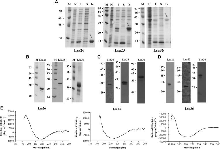
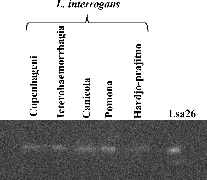
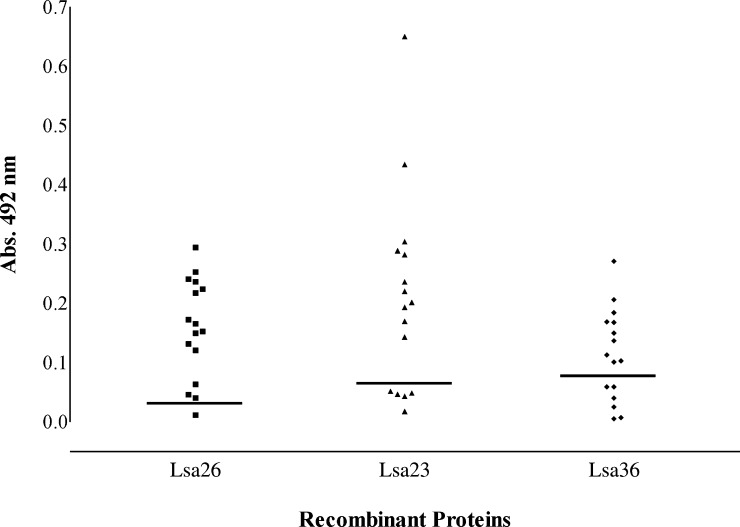
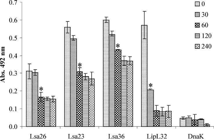
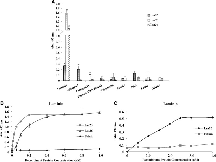
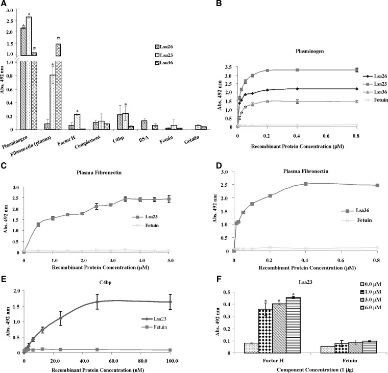
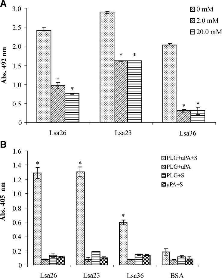
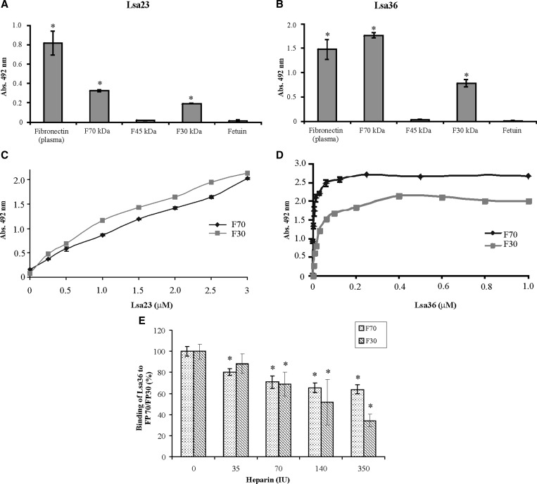
We report cloning, expression, purification, and characterization of three predicted leptospiral membrane proteins (LIC11360, LIC11009, and LIC11975). In silico analysis and proteinase K accessibility data suggest that these proteins might be surface exposed. We show that proteins encoded by LIC11360, LIC11009 and LIC11975 genes interact with laminin in a dose-dependent and saturable manner. The proteins are referred to as leptospiral surface adhesions 23, 26, and 36 (Lsa23, Lsa26, and Lsa36), respectively. These proteins also bind plasminogen and generate active plasmin. Attachment of Lsa23 and Lsa36 to fibronectin occurs through the involvement of the 30-kDa and 70-kDa heparin-binding domains of the ligand. Dose-dependent, specific-binding of Lsa23 to the complement regulator C4BP and to a lesser extent, to factor H, suggests that this protein may interfere with the complement cascade pathways. Leptospira spp. may use these interactions as possible mechanisms during the establishment of infection.


Figures

Figure 1.
Figure 1.
Analysis of LIC11009, LIC11360, and LIC11975 coding sequence conservation among strains of Leptospira spp. by Clustal W2 alignments. Blast analyses were assessed among sequences in GenBank, and leptospiral sequences were used to perform sequence alignment. Phylograms of all sequence alignments show the proximity of LIC11009 (A), LIC11360 (B), and LIC11975 (C) within pathogenic strains of Leptospira spp. The distant branches show the saprophytic strains.
Figure 2.
Figure 2.
Analysis of recombinant proteins of Leptospira interrogans by sodium dodecyl sulfate–polyacrylamide gel electrophoresis and staining with Coomassie blue, Western blotting, and circular dichroism. A, Lsa26, Lsa23, and Lsa36 expression from NaCl-induced Escherichia coli BL21-SI. M = molecular mass marker; NI = non-induced total bacterial extract; I = total bacterial cell lysates after induction; S = soluble fraction of the induced culture in the presence of 8 M urea; In = insoluble fraction of the induced culture. B, Purified recombinant proteins. C and D, Western blotting analyses of the recombinant proteins probed with monoclonal anti-His tag antibodies (1:1,000) (C) and correspondent antiserum produced in mice against each recombinant protein (diluted 1:5,000) (D). ECD spectra of Lsa26, Lsa23, and Lsa36 recombinant proteins are depicted after refolding. Far-ultraviolet CD spectra are shown as an average of five scans from 185 to 260 nm (E).Values on the left are in kilodaltons.
Figure 3.
Figure 3.
Protein distribution (Lsa26) among strains of Leptospira interrogans. Whole bacterial cell lysates and Lsa26 were separated by sodium dodecyl sulfate–polyacrylamide gel electrophoresis and transferred onto nitrocellulose membranes. Antiserum against recombinant Lsa26 was used as probe (diluted 1:50). Reactivity was shown by using an ECL reagent kit (GE Healthcare, Piscataway, NJ) and subsequent exposures with Gel Logic 2200 equipment (Equilab, Whitestone, NY). Recombinant protein Lsa26 was used as a marker.
Figure 4.
Figure 4.
Reactivity of recombinant proteins Lsa26, Lsa23 and Lsa36 of Leptospira interrogans with serum samples of persons given a diagnosis of leptospirosis. Positive serum samples (responders) were determined by using an enzyme-linked immunosorbent assay with recombinant proteins and serum samples from patients. Reactivity was evaluated as total IgG. The cutoff value (horizontal lines) for each recombinant protein was defined as the absorbance (Abs.) value for the 96th percentile of two commercial pools of healthy human serum.
Figure 5.
Figure 5.
Protease accessibility assay of LIC11009, LIC11360 and LIC11975 coding sequences of Leptospira interrogans. Viable L. interrogans serovar Copenhageni strain M-20 were incubated with 25 μg/mL of proteinase K at the indicated times (in minutes). Suspensions were centrifuged, washed, resuspended in phosphate-buffered saline, coated onto a microplate. Antibodies against Lsa26, Lsa23 Lsa36 were used. Antiserum against the outer membrane protein LipL32 and the cytoplasmic leptospiral protein DnaK were used as positive and negative controls, respectively. Parallel blanks were run without antibodies against the recombinant proteins. Bars represent the mean ± SD absorbance of three replicates for each protein and are representative of two independent experiments. For statistical analyses, the optical density value after treatment with proteinase K was compared with the value at 0 hours incubation by a two-tailed t-test (*P < 0.05).
Figure 6.
Figure 6.
Interaction of recombinant proteins of Leptospira interrogans with extracellular matrix (ECM) components. A, One microgram of ECM macromolecules was coated onto enzyme-linked immunosorbent assay (ELISA) microplates wells after incubation with 1 μg of Lsa26, Lsa23, or Lsa36. Bovine serum albumin, gelatin, and fetuin were used in the place of ECM as negative controls for nonspecific binding. Binding was evaluated by ELISA. Bars represent the mean ± SD absorbance at 492 nm of three replicates for each protein and are representative of two independent experiments. For statistical analyses, the interaction of recombinant proteins with ECM was compared with its binding to fetuin by two-tailed t-test (*P < 0.05). B and C, Lsa23 and Lsa36, and Lsa26 dose-dependent binding experiments with laminin, respectively. Binding was detected by polyclonal antibodies against each recombinant protein; fetuin was included as a negative control. Dissociation constant data are shown in Table 2.
Figure 7.
Figure 7.
Attachment of Lsa26, Lsa23 and Lsa36 proteins of Leptospira interrogans to plasma components. A, Wells were coated with 1 μg of each plasma component or control proteins bovine serum albumin, gelatin, and fetuin followed by incubation with 1 μg of recombinant proteins per well. Binding was measured by enzyme-linked immunosorbent assay (ELISA). Data represent the mean ± SD absorbance at 492 nm of three replicates for each protein and are representative of two independent experiments. For statistical analyses, the attachment of recombinant proteins to plasma components was compared with its binding to fetuin by two-tailed t-test (*P < 0.05). Proteins that showed reactivity were further assayed with the respective component. Dose-dependent binding experiments with plasminogen (B), plasma fibronectin (C and D), C4BP (E), and with factor H (F). One microgram of each plasma component was immobilized onto 96-well ELISA plates and increasing concentrations of each recombinant protein were added. Binding was detected by using antiserum raised in mice against each recombinant protein at an appropriate dilution. Fetuin was included as a negative control. Each experiment was performed in triplicate and expressed as the mean ± SD absorbance at 492 nm for each point and are representative of two independent experiments. Calculated equilibrium constants are shown in Table 2. Attachment of Lsa23 with factor H was compared with its binding to fetuin by two-tailed t-test (*P < 0.05).
Figure 8.
Figure 8.
Role of lysine residues in recombinant protein–plasminogen (PLG) interaction and generation of plasmin (PLA) in Leptospira interrogans. A, Binding of Lsa26, Lsa23, and Lsa36 (10 μg/mL) to PLG was conducted in the presence or absence (no inhibition) of the lysine analog 6-aminocaproic acid (ACA). Bound PLG was detected and quantified by specific antibodies for each recombinant protein. Bars represent the mean ± SD absorbance at 492 nm of three replicates and are representative of two independent experiments. Attachment of recombinant protein in the presence of ACA was compared with its binding to PLG in the absence of ACA by two-tailed t-test (*P < 0.05). B, PLA generation by PLG bound to recombinant proteins was assayed by using a modified enzyme-linked immunosorbent assay. Immobilized recombinant proteins were treated as follows: PLG + urokinase-type PLG activator [uPA] + specific PLA substrate (PLG + uPA + S) or controls lacking one of the three components (PLG + uPA; PLG + S; uPA + S). Bovine serum albumin (BSA) was used as a negative control. Bars represent mean ± SD absorbance at 405 nm as a measure of relative substrate degradation of three replicates for each condition and are representative of two independent experiments. Statistically significant binding was calculated in comparison with the negative control (BSA) (*P < 0.05).
Figure 9.
Figure 9.
Binding of Lsa23 and Lsa36 of Leptospira interrogans with plasma fibronectin proteolytic fragments. One microgram of proteolytic fragments F70, F45, and F30 were coated onto microtiter plates followed by incubation with 1 μg of (A) Lsa23 or (B) Lsa36. Fetuin was used as negative control for nonspecific binding. Bars represent the mean ± SD absorbance at 492 nm of three replicates for each protein and are representative of two independent experiments; the attachment of recombinant proteins to proteolytic fragments was compared with its binding to fetuin by two-tailed t-test (*P < 0.005). C and D, dose-dependent binding experiments of Lsa23 and Lsa36, respectively, with F70 and F30; each experiment was performed in triplicate and results expressed as the mean ± SD absorbance 492 nm for each point. Dissociation constants are shown in Table 2. E, Inhibition of Lsa36 interaction with F70 and F30 by heparin. Attachment of Lsa36 (1 μg) to fixed F70 and F30 concentration was performed in the presence of increasing amounts of heparin (0–350 IU). Attachment of Lsa36 in the presence of heparin was compared with its binding to each proteolytic fragments without heparin (0 IU) by two-tailed t-test (*P < 0.05).

Similar articles

Cited by

References

    1. Bharti AR, Nally JE, Ricaldi JN, Matthias MA, Diaz MM, Lovett MA, Levett PN, Gilman RH, Willig MR, Gotuzzo E, Vinetz JM. Leptospirosis: a zoonotic disease of global importance. Lancet Infect Dis. 2003;3:757–771. - PubMed
    1. de la Pena-Moctezuma A, Bulach DM, Adler B Genetic differences among the LPS biosynthetic loci of serovars of Leptospira interrogans and Leptospira borgpetersenii. FEMS Immunol Med Microbiol. 2001;31:73–81. - PubMed
    1. Faine S, Adler B, Bolin C, Perolat P. Leptospira and Leptospirosis. Melbourne. Australia: MediSci Press; 1999.
    1. Nakai K, Kanehisa M. Expert system for predicting protein localization sites in gram-negative bacteria. Proteins. 1991;11:95–110. - PubMed
    1. Juncker AS, Willenbrock H, Von Heijne G, Brunak S, Nielsen H, Krogh A. Prediction of lipoprotein signal peptides in gram-negative bacteria. Protein Sci. 2003;12:1652–1662. - PMC - PubMed

Publication types

MeSH terms

LinkOut - more resources