. 2013 Oct 1;4(7):612-23.
doi: 10.4161/viru.26055. Epub 2013 Aug 13.

The non-structural (NS) gene segment of H9N2 influenza virus isolated from backyard poultry in Pakistan reveals strong genetic and functional similarities to the NS gene of highly pathogenic H5N1

Affiliations

The non-structural (NS) gene segment of H9N2 influenza virus isolated from backyard poultry in Pakistan reveals strong genetic and functional similarities to the NS gene of highly pathogenic H5N1

Muhammad Munir et al. Virulence. .

Abstract

Apart from natural reassortment, co-circulation of different avian influenza virus strains in poultry populations can lead to generation of novel variants and reassortant viruses. In this report, we studied the genetics and functions of a reassorted non-structural gene (NS) of H9N2 influenza virus collected from back yard poultry (BYP) flock. Phylogenetic reconstruction based on hemagglutinin and neuraminidase genes indicates that an isolate from BYP belongs to H9N2. However, the NS gene-segment of this isolate cluster into genotype Z, clade 2.2 of the highly pathogenic H5N1. The NS gene plays essential roles in the host-adaptation, cell-tropism, and virulence of influenza viruses. However, such interpretations have not been investigated in naturally recombinant H9N2 viruses. Therefore, we compared the NS1 protein of H9N2 (H9N2/NS1) and highly pathogenic H5N1 (H5N1/NS1) in parallel for their abilities to regulate different signaling pathways, and investigated the molecular mechanisms of IFN-β production in human, avian, and mink lung cells. We found that H9N2/NS1 and H5N1/NS1 are comparably similar in inhibiting TNF-α induced nuclear factor κB and double stranded RNA induced activator protein 1 and interferon regulatory factor 3 transcription factors. Thus, the production of IFN-β was inhibited equally by both NS1s as demonstrated by IFN stimulatory response element and IFN-β promoter activation. Moreover, both NS1s predominantly localized in the nucleus when transfected to human A549 cells. This study therefore suggests the possible increased virulence of natural reassortant viruses for their efficient invasion of host immune responses, and proposes that these should not be overlooked for their epizootic and zoonotic potential.

Keywords: IFN production; NS1; immune regulation; influenza; reassortment.


Figures

None
Figure 1. Phylogenetic relationships of the (A) HA, (B) NA, and (C) NS genes of representative influenza A viruses isolated in Asia. Trees were generated by the neighbor-joining method with the MEGA program (Bayesian analysis revealed similar relationships). Numbers above the branches indicate neighbor-joining bootstrap values. Only bootstrap values above 80% are shown. The analysis was based on nucleotides 129 to 1042 of the HA gene, 231 to 1297 of the NA gene and 88 to 815 of the NS gene. The isolate characterized in this study is labeled with a black square (◼) whereas previously characterized isolates from Pakistan are labeled with a black circle (●). The Swedish isolate used in this study is marked with a star (★) (C). An alternative genotype characterization is shown in brackets. A bar represents 0.01–0.05 substitutions per site.
None
Figure 2. Inhibition of dsRNA or TNF-α induced pathways by overexpression of the NS1 protein of influenza A viruses. A 500 ng of expression plasmids for NS1 protein derived from H9N2 (H9N2/NS1) and H5N1 (H5N1/NS1) were cotransfected with 10 ng of pGL4.74 together with 500 ng of (A) 4×IRF3-Luc, (B) pAP-1-Luc, and (C) NFκB-Luc reporter plasmids or normalized with empty flag vector (cont.) or left untreated (cells). After 24 h post-transfection, the A549 cells were either stimulated with 10 μg of dsRNA (poly I:C) per ml (4×IRF3 and pAP-1) or 25 ng/ml of TNF-α (NFκB) or left untreated (cells). After an additional 24 h of incubation period, the cell extracts were prepared (A–C). CEF cell were transfected with corresponding constructs, and all the conditions were maintained as practiced for A549 cells. After 24 h of stimulation of (D) 4×IRF3 and (E) pAP-1 promoter with (F) 10 μg of dsRNA per ml or NFκB promoter with 25 ng/ml of TNF-α or untreated cells, the luciferase extracts were prepared. In both cell lines, the luciferase activity was measured using the Dual-Luciferase Assay System (Promega). The values in the mock-transfected cells were normalized and were set to 100% (4×IRF3 cont., pAP-1 cont., and NFκB cont.). Error bars indicate standard deviations. The data shown here are representative for 3 experiments with transfections performed in duplicate. *indicates a significant difference as determined by the Student t test, with P values of <0.05.
None
Figure 3. Expression patterns of H9N2/NS1 and H5N1/NS1 proteins in subcellular compartments in A549. (A) The A549 cells were transfected with 500 ng of expression plasmids encoding NS1 from H9N2/NS1 and H5N1/NS1 or with empty flag vector (mock treated). After 24 h post-transfection, the cells were lysed and subjected to western blotting by stained with anti-flag and anti-β-actin antibodies. (B) Transfections were performed as demonstrated for western blotting. After 18 h of transfection, cells were fixed and processed for in situ PLA. The images reflect a predominant expression of NS1 proteins in the nucleus. Both H9N2/NS1 and H5N1/NS1 proteins showed comparable expression as was seen in the western blotting. The images were taken at 20×.
None
Figure 4. Inhibition of pre- and mature-IFN-β mRNA transcription in the presence of H9N2/NS1 and H5N1/NS1 proteins. A549 cells were transfected with H9N2/NS1 and H5N1/NS1 expression plasmids or left untreated (poly I:C stimulated and cells). After 24 h of post-transfection incubation period, the cells were either left unstimulated (cells) or stimulated with 10 μg of dsRNA per ml. Cells were TRIzole lysed and RNA was extracted at 0, 2, 4, 8, 16, and 24 h post-stimulation. Equal amounts of RNA from all time points were used to synthesis cDNA for (A) pre-mRNA (with 20 mer oligonucleotide) and for (B) mature-mRNA (with poly dT[20] primer). Both cDNAs were subjected to real-time PCR for IFN-β and β-actin mRNA quantification. The results were presented as fold increase in IFN-β mRNA using the 2-ΔΔT method. *Indicates significant differences at P < 0.05 (the Student t test) in 3 independent experiments.
None
Figure 5. The H9N2/NS1 and H5N1/NS1 proteins inhibit the IFN-β gene expression in VSV-GFP bioassay. (A) The stimulation of IFN-β by dsRNA is inhibited in the presence of NS1 proteins and thus leads to efficient replication of VSV-GFP virus. (B) A549 cells were transfected with 300 ng of expression plasmids encoding H9N2/NS1 and H5N1/NS1 proteins or left untreated (mock treated). After 18 h post-transfection incubation period, the cells were stimulated with 5 μg of dsRNA per ml. After an additional 24 h, the cells were infected with VSV-GFP (MOI of 2). Fluorescence was measured after 18 h of infection. The images were taken at 20×. The quantitative analysis of the infected cells effectively expressing replication of VSV-GFP in the presence of NS1 protein from both H5N1 and H9N2. Bars represent a non-significant difference at P > 0.05 (the Student t test) in 3 independent experiments.
None
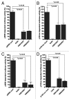
Figure 6. Inhibition of dsRNA induced ISRE and IFN-β promoter activities by overexpression of NS1 protein of influenza A viruses. A 500 ng of expression plasmids for NS1 protein derived from H9N2 (H9N2/NS1) and H5N1 (H5N1/NS1) were cotransfected with 10 ng of pGL4.74 together with 500 ng of (A) pISRE-Luc, (B) p125-Luc reporter plasmids, or normalized with empty flag vector (cont.) or left untreated (cells). After 24 h post-transfection, the A549 cells were either stimulated with 10 μg of dsRNA (poly I:C) per ml or not stimulated (cells). After an additional 24 h of incubation period, the cell extracts were prepared (A and B). CEF cell were transfected with corresponding constructs and all the conditions were maintained as practiced for A549 cells. After 24 h of stimulation of (C) pISRE-Luc, (D) p125-Luc reporter plasmids with 10 μg of dsRNA per ml or untreated cells, and the luciferase extracts were prepared. In both cell lines, the luciferase activity was measured using the Dual-Luciferase Assay System (Promega). The values in mock-transfected cells were normalized and were set to 100% (pISRE cont. and p125-Luc cont.). Error bars indicate standard deviations. The data shown here are representative for three experiments with transfections performed in duplicate. *indicates a significant difference as determined by the Student t test, with P value of <0.05.
None
Figure 7. The NS1 proteins of H9N2 and H5N1 inhibit dsRNA induced nuclear localization of IRF3. A549 cells were transfected with 300 ng of NS1 expression constructs for both H9N2 and H5N1 or mock transfected. After 24 h of incubation, cells were stimulated with 10 μg dsRNA per ml and after additional 24 h cells were fixed and probed with pIRF3 (ser396) antibodies. The images were taken at 20×.

Comment in

References

    1. Homme PJ, Easterday BC. Avian influenza virus infections. I. Characteristics of influenza A-turkey-Wisconsin-1966 virus. Avian Dis. 1970;14:66–74. doi: 10.2307/1588557. - DOI - PubMed
    1. Guan Y, Shortridge KF, Krauss S, Webster RG. Molecular characterization of H9N2 influenza viruses: were they the donors of the “internal” genes of H5N1 viruses in Hong Kong? Proc Natl Acad Sci U S A. 1999;96:9363–7. doi: 10.1073/pnas.96.16.9363. - DOI - PMC - PubMed
    1. Cong YL, Wang CF, Yan CM, Peng JS, Jiang ZL, Liu JH. Swine infection with H9N2 influenza viruses in China in 2004. Virus Genes. 2008;36:461–9. doi: 10.1007/s11262-008-0227-z. - DOI - PubMed
    1. Peiris M, Yam WC, Chan KH, Ghose P, Shortridge KF. Influenza A H9N2: aspects of laboratory diagnosis. J Clin Microbiol. 1999;37:3426–7. - PMC - PubMed
    1. Lin YP, Shaw M, Gregory V, Cameron K, Lim W, Klimov A, Subbarao K, Guan Y, Krauss S, Shortridge K, et al. Avian-to-human transmission of H9N2 subtype influenza A viruses: relationship between H9N2 and H5N1 human isolates. Proc Natl Acad Sci U S A. 2000;97:9654–8. doi: 10.1073/pnas.160270697. - DOI - PMC - PubMed

Publication types

MeSH terms

LinkOut - more resources