. 2013 Dec;21(12):2195-204.
doi: 10.1038/mt.2013.191. Epub 2013 Aug 20.

Proapoptotic peptide-mediated cancer therapy targeted to cell surface p32

Affiliations

Proapoptotic peptide-mediated cancer therapy targeted to cell surface p32

Lilach Agemy et al. Mol Ther. 2013 Dec.

Erratum in

  • Mol Ther. 2014 Nov;22(11):2013

Abstract

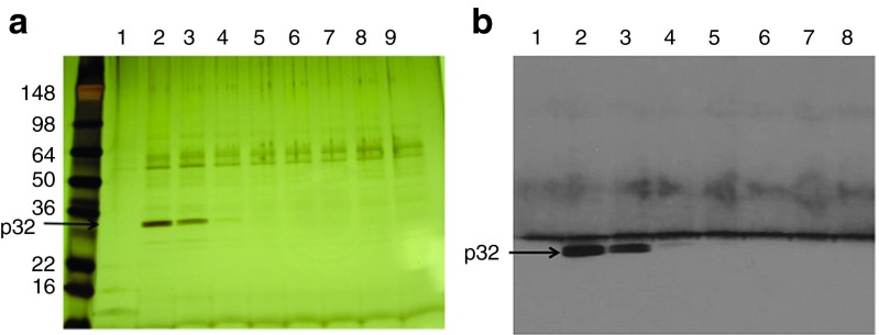
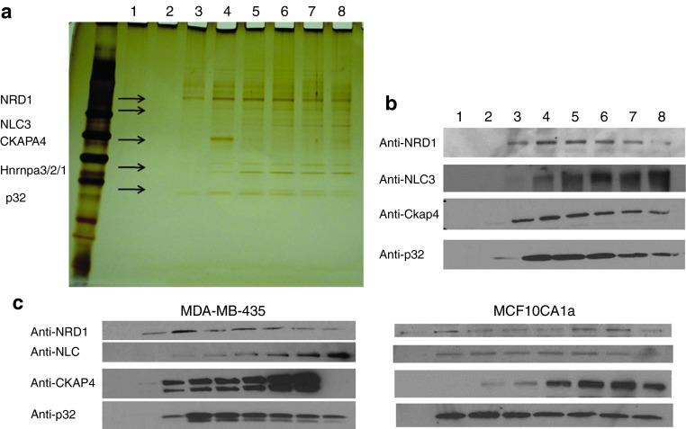
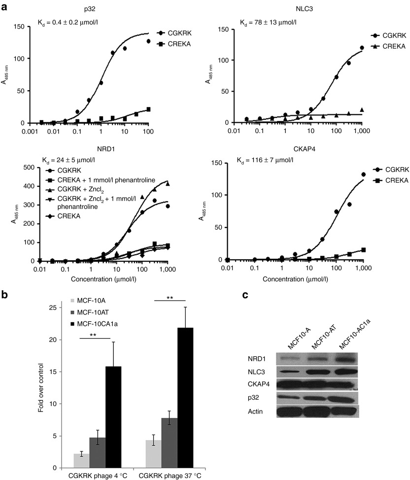
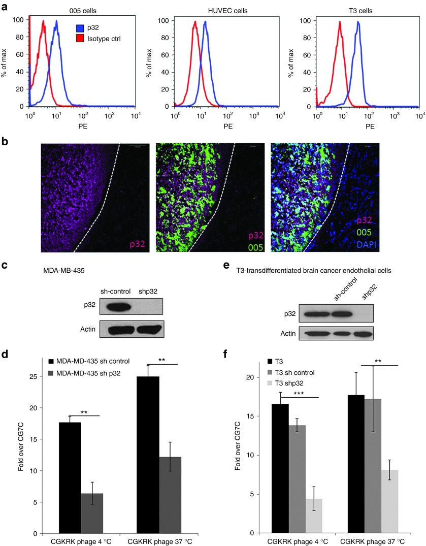
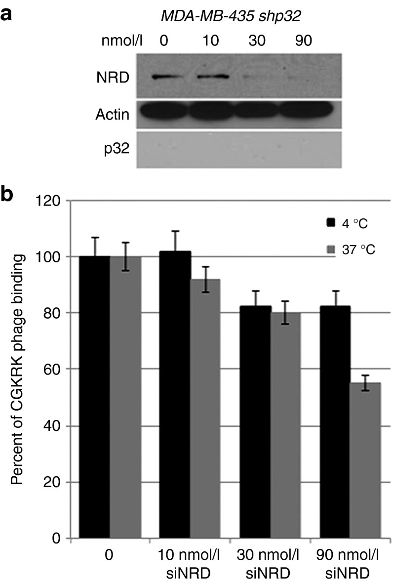
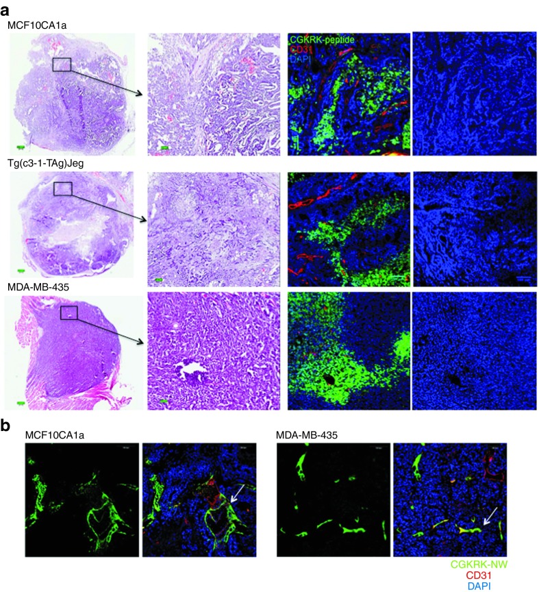
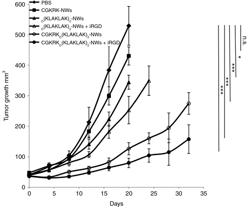
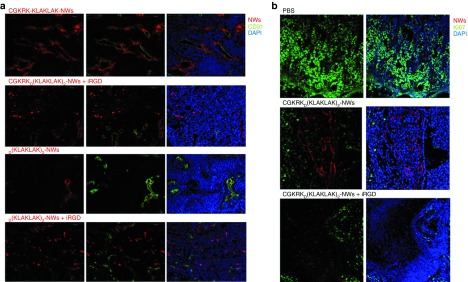
Antiangiogenic therapy is a promising new treatment modality for cancer, but it generally produces only transient tumor regression. We have previously devised a tumor-targeted nanosystem, in which a pentapeptide, CGKRK, delivers a proapoptotic peptide into the mitochondria of tumor blood vessel endothelial cells and tumor cells. The treatment was highly effective in glioblastoma mouse models completely refractory to other antiangiogenic treatments. Here, we identify p32/gC1qR/HABP, a mitochondrial protein that is also expressed at the cell surface of activated (angiogenic) endothelial cells and tumor cells, as a receptor for the CGKRK peptide. The results demonstrate the ability of p32 to cause internalization of a payload bound to p32 into the cytoplasm. We also show that nardilysin, a protease capable of cleaving CGKRK, plays a role in the internalization of a p32-bound payload. As p32 is overexpressed and surface displayed in breast cancers, we studied the efficacy of the nanosystem in this cancer. We show highly significant treatment results in an orthotopic model of breast cancer. The specificity of cell surface p32 for tumor-associated cells, its ability to carry payloads to mitochondria, and the efficacy of the system in important types of cancer make the nanosystem a promising candidate for further development.


Figures

Figure 1
Figure 1
Identification of a CGKRK-binding protein in mitochondrial protein extracts. Protein extracts prepared from mouse liver mitochondria were fractionated by affinity chromatography on CGKRK-coupled columns. Bound proteins were eluted with 2 mmol/l CGKRK peptide solution and separated by sodium dodecyl sulfate–polyacrylamide gel electrophoresis. (a) Silver-stained gel. The arrow points to the band identified as p32 by mass spectrometry, and (b) immunoblotting detection of p32.
Figure 2
Figure 2
Identification of CGKRK-binding proteins in brain tumor protein extracts. Protein extracts prepared from 005 mouse brain tumors were fractionated on CGKRK affinity columns. Bound proteins were eluted with 2 mmol/l CGKRK peptide solution and separated by sodium dodecyl sulfate–polyacrylamide gel electrophoresis. (a) Silver-stained gel. The arrows point to the bands subjected to mass spectrometry for protein identification. The proteins identified are shown on the left side of the arrows. (b) Immunoblot with anti-p32, anti-CKAP4, anti-nucleolin, and anti-nardilysin (NRD). (c) Protein extracts prepared from MDA-MB-435 and MCF10CA1a tumors were fractionated on a CGKRK affinity column. Bound proteins were eluted with 2 mmol/l CGKRK solution. Results of immunoblotting with anti-p32, anti-CKAP4, anti-nucleolin, and anti-NRD are shown.
Figure 3
Figure 3
CGKRK binding to candidate receptor proteins. (a) Binding of FAM–CGKRK to purified p32, NLC3, CKAP4, and nardilysin1 (NRD1). Binding of increasing amounts of FAM–CGKRK peptide and a control FAM–CREKA peptide to immobilized p32, NLC3, CKAP4, and NRD1 proteins was detected by fluorescence and normalized to nonspecific binding to the plastic plate. One representative binding curve out of three independent experiments with similar results is shown. The Kd was calculated from all three experiments using Prism software. (b) Binding and internalization of CGKRK phage particles in a series of MCF10 human breast tumor cells. MCF10 cells were incubated with phage displaying CGKRK or a polyglycine control peptide, CG7C, overnight at 4 °C or for 1 hour at 37 °C. To assess internalization, phages bound at the cell surface were removed by washing the cells with an acid buffer before phage titration. Statistical analyses were performed with Student's t-test. n = 3; error bars, mean ± SEM; **P < 0.01. (c) Total expression levels of NRD-1, NLC3, CKAP4, and p32 in the MCF10 cells assessed by immunoblotting.
Figure 4
Figure 4
p32-dependent binding of CGKRK to tumor cells. (a) Cell-surface expression of p32 on 005 brain tumor cells, T3 brain tumor endothelial cells, and human umbilical vein endothelial cells assessed by flow cytometry. (b) Localization of p32 in 005 brain tumors. Frozen sections of mouse brain containing green fluorescent protein–expressing 005 brain tumor cells (green) were stained for p32 (magenta) and nuclei (blue) and were imaged under a confocal microscope. Scale bars, 100 µm. (c–f) Effect of p32 knockdown on CGKRK binding. p32 expression levels were determined by immunoblot on whole-cell lysates from MDA-MB-435 cells (c) or T3 cells (e) stably expressing p32 short hairpin RNA (shRNA) or control shRNA. β-actin was used as a loading control. Cells with varying p32 expression levels were incubated with phages displaying CGKRK or CG7C overnight at 4 °C or for 1 hour at 37 °C. To assess internalization, phages bound at the cell surface were removed by washing the cells with an acid buffer before phage titration. Note that the p32 knockdown resulted in reduced CGKRK binding. Statistical analysis was performed with Student's t-test;**P < 0.01; ***P < 0.001; n = 3; error bars, mean ± SEM.
Figure 5
Figure 5
Effect of nardilysin (NRD) knockdown on CGKRK phage binding and internalization into cells. (a) MDA-MB-435 cells were transiently transfected with NRD small interfering RNA (siRNA) or control siRNA. After 48 hours, NRD expression levels were determined by immunoblotting. β-actin was used as a loading control. The results are representative of three independent experiments. (b) The cells transfected with NRD siRNA or control siRNA were incubated with CGKRK phage for 1 hour at 37 °C to assess internalization into cells or overnight at 4 °C to assess cell binding. In internalization assays, the phages bound at the cell surface were removed by washing the cells with an acid buffer before phage titration. n = 3; error bars, mean ± SEM.
Figure 6
Figure 6
Homing of CGKRK peptide and CGKRK-nanoworms (NWs) to breast tumors. Mice bearing MCF10CA1a, MDA-MB-435, or MMTV-PyMT orthotopic tumors were intravenously injected with (a) 200 µg of FAM-CGKRK peptide or (b) 5 mg iron/kg of FAM-CRGDK-coated NWs. Circulation time was 3 hours for the peptide and 5–6 hours for the NWs. The mice were perfused through the heart with PBS, and tissues were collected and processed for immunofluorescence. Representative confocal microscopy images from three mice per tumor type are provided. Arrows in panel b point to tumor blood vessels targeted by CGKRK-NW. Green, (a) FAM-CGKRK and (b) CGKRK-NWs; red, CD31; blue, nuclei; Scale bars, (a) 200 μm and (b) 100 μm.
Figure 7
Figure 7
CGKRKD[KLAKLAK]2-nanoworm (NW) treatment in MCF10CA1a tumor mice. Mice bearing MCF10CA1a orthotopic tumor xenografts were intravenously treated with peptide-coated NWs every other day for 3 weeks at a dose of 5 mg/kg. In some groups, 100 µg of iRGD was coinjected with the NWs to enhance extravasation and tissue penetration of the particles. PBS, n = 7; CGKRK-NW, n = 5;D[KLAKLAK]2-NWs, n = 6; [KLAKLAK]2-NWs+iRGD, n = 6; CGKRKD[KLAKLAK]2-NWs, n = 8; CGKRKD[KLAKLAK]2-NWs+iRGD, n = 8. a, Tumor growth was followed throughout the treatment and for an additional 14 days posttreatment period in the groups, where the size of the tumors stayed below the size limit allowed by the institutional review committee (in the Materials and Methods section). The experiment was done twice. Error bars, mean ± SD. Statistical analyses were performed with ANOVA; n.s., not significant; *P < 0.05; ***P < 0.001.
Figure 8
Figure 8
Nanoworm (NW) distribution and Ki67 staining in tumors of treated mice. At the end of a 3-week treatment of MCF10CA1a tumor in a study similar the one shown in Figure 7, the tumors were collected and processed for immunofluorescence. Confocal microscopy images of tumors from four representative animals in each group are shown. Red, NWs; green, (a) CD31 and (b) Ki67; blue, nuclei. Scale bars, 100 μm.

References

    1. Ferrari M. Frontiers in cancer nanomedicine: directing mass transport through biological barriers. Trends Biotechnol. 2010;28:181–188. - PMC - PubMed
    1. Ferrara N, Alitalo K. Clinical applications of angiogenic growth factors and their inhibitors. Nat Med. 1999;5:1359–1364. - PubMed
    1. Ruoslahti E. Peptides as targeting elements and tissue penetration devices for nanoparticles. Adv Mater Weinheim. 2012;24:3747–3756. - PMC - PubMed
    1. Gradishar WJ, Tjulandin S, Davidson N, Shaw H, Desai N, Bhar P, et al. Phase III trial of nanoparticle albumin-bound paclitaxel compared with polyethylated castor oil-based paclitaxel in women with breast cancer. J Clin Oncol. 2005;23:7794–7803. - PubMed
    1. Haley B, Frenkel E. Nanoparticles for drug delivery in cancer treatment. Urol Oncol. 2008;26:57–64. - PubMed

Publication types

MeSH terms