Review
. 2013 Sep 6;219(1):R1-R12.
doi: 10.1530/JOE-13-0276. Print 2013 Oct.

Cartilage to bone transitions in health and disease

Affiliations
Review

Cartilage to bone transitions in health and disease

K A Staines et al. J Endocrinol. .

Abstract

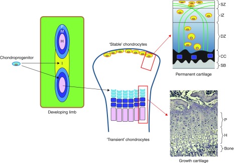
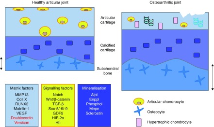
Aberrant redeployment of the 'transient' events responsible for bone development and postnatal longitudinal growth has been reported in some diseases in what is otherwise inherently 'stable' cartilage. Lessons may be learnt from the molecular mechanisms underpinning transient chondrocyte differentiation and function, and their application may better identify disease aetiology. Here, we review the current evidence supporting this possibility. We firstly outline endochondral ossification and the cellular and physiological mechanisms by which it is controlled in the postnatal growth plate. We then compare the biology of these transient cartilaginous structures to the inherently stable articular cartilage. Finally, we highlight specific scenarios in which the redeployment of these embryonic processes may contribute to disease development, with the foresight that deciphering those mechanisms regulating pathological changes and loss of cartilage stability will aid future research into effective disease-modifying therapies.

Keywords: bone; cartilage; chondrocyte; endochondral ossification; osteoarthritis.


Figures

Figure 1
Figure 1
Schematic representation depicting the proposed developmental origins of articular and growth plate cartilage. Mesenchymal aggregation of chondro-progenitors forms the cartilage anlagen and stages of chondrocyte proliferation (P, light blue), maturation (M, purple) and hypertrophy (H, pink) emerge to provide the origins of the future ‘transient’ chondrocytes of the growth plate cartilage. Intervening regions of progressively condensing mesenchyme define the interzone (I, yellow) regions; the position of the future joint and origins of the ‘stable’ articular cartilage chondrocytes. Images (right hand side) depicting the organisation of the mature growth plate (lower) and articular cartilage (upper), consisting of uncalcified cartilage zones (superficial (SZ), intermediate (IZ) and deep (DZ)), and the related chondrocyte and collagen fibril arrangement. The tidemark separates the non-calcified cartilage from the calcified cartilage (CC) which overlies the subchondral bone (SB). Bar=0.1 mm.
Figure 2
Figure 2
Schematic diagram depicting the healthy articular joint and the osteoarthritic joint in which articular cartilage fibrillation and degradation is observed with concomitant subchondral bone thickening. Contributing to this osteoarthritic pathology, the normally ‘stable’ articular chondrocytes of the articular cartilage adopt a ‘transient’ phenotype with observed chondrocyte hypertrophy and matrix mineralisation similar to that seen in the growth cartilage depicted in Fig. 1. Potential regulation of these processes may include changes in the expression of matrix factors (blue box; factors induced in osteoarthritis development in black, factors lost in red), signalling pathways affecting chondrocyte phenotype and function (yellow box) and the known regulators of mineralisation processes (purple box) (Grover & Roughley 1993, Pacifici et al. 2006, Zhang et al. 2007, Fosang & Beier 2011, Pitsillides & Beier 2011, Staines et al. 2012b). Lessons may be learnt from these and their application may better identify disease aetiology.

References

    1. Abad V, Meyers JL, Weise M, Gafni RI, Barnes KM, Nilsson O, Bacher JD, Baron J. The role of the resting zone in growth plate chondrogenesis. Endocrinology. 2002;143:1851–1857. doi: 10.1210/en.143.5.1851. - DOI - PubMed
    1. Adams CS, Shapiro IM. The fate of the terminally differentiated chondrocyte: evidence for microenvironmental regulation of chondrocyte apoptosis. Critical Reviews in Oral Biology and Medicine. 2002;13:465–473. doi: 10.1177/154411130201300604. - DOI - PubMed
    1. Ahmed SF, Farquharson C. The effect of GH and IGF1 on linear growth and skeletal development and their modulation by SOCS proteins. Journal of Endocrinology. 2010;206:249–259. doi: 10.1677/JOE-10-0045. - DOI - PubMed
    1. Aigner T, Reichenberger E, Bertling W, Kirsch T, Stoss H, von der Mark K. Type X collagen expression in osteoarthritic and rheumatoid articular cartilage. Virchows Archiv. B, Cell Pathology Including Molecular Pathology. 1993;63:205–211. doi: 10.1007/BF02899263. - DOI - PubMed
    1. Amling M, Neff L, Tanaka S, Inoue D, Kuida K, Weir E, Philbrick WM, Broadus AE, Baron R. Bcl-2 lies downstream of parathyroid hormone-related peptide in a signaling pathway that regulates chondrocyte maturation during skeletal development. Journal of Cell Biology. 1997;136:205–213. doi: 10.1083/jcb.136.1.205. - DOI - PMC - PubMed

Publication types