Review
. 2013 Sep;37(9):1683-95.
doi: 10.1007/s00264-013-2030-2.

Supramalleolar osteotomies for degenerative joint disease of the ankle joint: indication, technique and results

Affiliations
Review

Supramalleolar osteotomies for degenerative joint disease of the ankle joint: indication, technique and results

Alexej Barg et al. Int Orthop. 2013 Sep.

Abstract

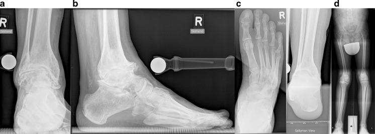
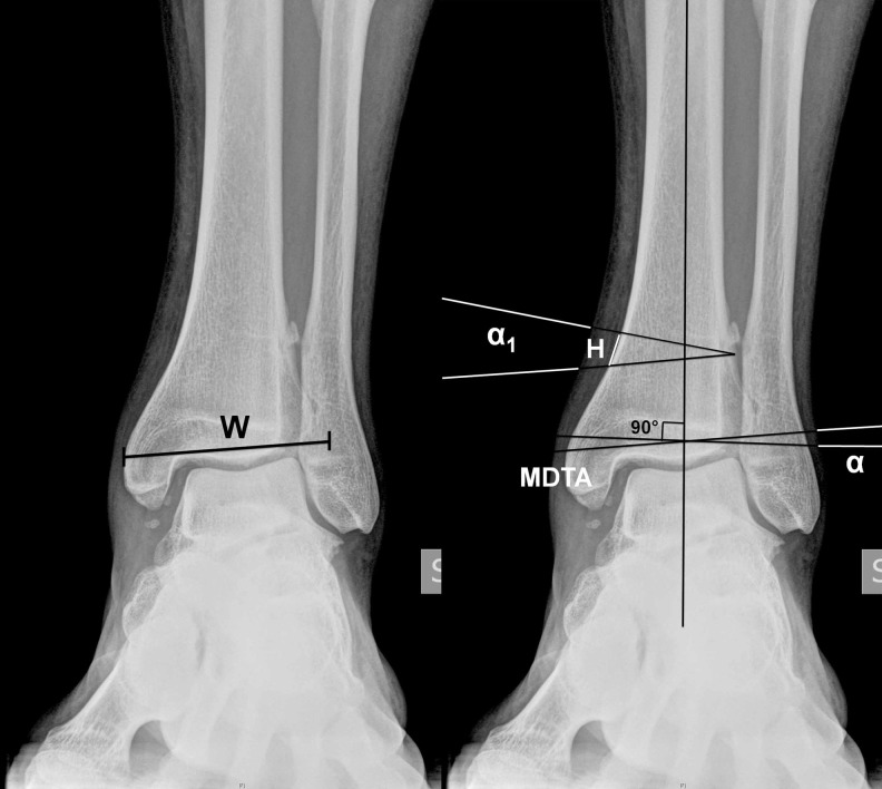
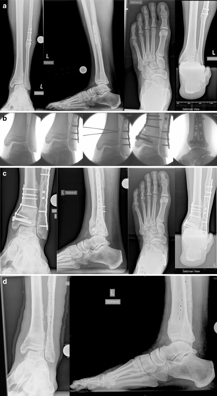
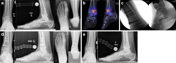
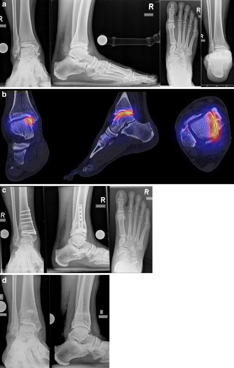
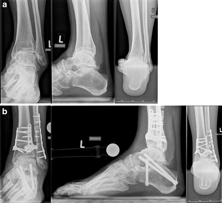
Patients with varus or valgus hindfoot deformities usually present with asymmetric ankle osteoarthritis. In-vitro biomechanical studies have shown that varus or valgus hindfoot deformity may lead to altered load distribution in the tibiotalar joint which may result in medial (varus) or lateral (valgus) tibiotalar joint degeneration in the short or medium term. The treatment of asymmetric ankle osteoarthritis remains challenging, because more than half of the tibiotalar joint surface is usually preserved. Therefore, joint-sacrificing procedures like total ankle replacement or ankle arthrodesis may not be the most appropriate treatment options. The shortand midterm results following realignment surgery, are very promising with substantial pain relief and functional improvement observed post-operatively. In this review article we describe the indications, surgical techniques, and results from of realignment surgery of the ankle joint in the current literature.


Figures

Fig. 1
Fig. 1
Radiographic assessment of foot and ankle. Radiographic evaluation of affected ankles with weight-bearing radiographs, including anteroposterior view of the ankle (a), lateral (b) and dorsoplantar (c) views of the ankle, and Saltzman view of the hindfoot (d). Whole leg radiograph is used to assess concomitant deformities of the lower extremity (d)
Fig. 2
Fig. 2
Pre-operative planning. Pre-operative weight-bearing anteroposterior radiograph is used for the planning of a supramalleolar medial closing wedge osteotomy. aW width of the distal part of the tibia (in this case 62 mm). bMDTA medial distal tibial angle (in this case 97.5°), α valgus deformity (in this case 97.5°), α1 amount of valgus deformity with desired overcorrection (in this case 7.5 + 2° = 9.5°), H height of the wedge to be removed (in this case tan 9.5° × 62 mm = 10 mm)
Fig. 3
Fig. 3
Medial closing wedge osteotomy and corrective Z-shaped osteotomy of the fibula. a Pre-operative weight-bearing radiographs show post-traumatic valgus tilting of the talus within the mortise and malunion of the fibula with substantial shortening and external rotation. Saltzman view shows the valgus hindfoot alignment. b First, corrective Z-shaped osteotomy of the fibula was performed to achieve elongation of the fibula and derotation as well as an aligned ankle mortise. Then medial closing wedge osteotomy was performed to address the valgus hindfoot deformity. c Post-operative weight-bearing radiographs show completed osseous healing at the site of osteotomies at the 1-year follow-up. Saltzman view shows normal hindfoot alignment. d After hardware removal patient is pain-free with no restrictions of sports activities
Fig. 4
Fig. 4
Medial closing wedge osteotomy and lateral lengthening calcaneal osteotomy. a Pre-operative weight-bearing radiographs show incipient degenerative changes of the lateral tibiotalar joint with slight valgus tilting of the talus within the mortise. Saltzman view shows the valgus hindfoot alignment. b SPECT/CT shows biologically active subchondral cysts in the lateral tibiotalar joint. c Supramalleolar medial closing wedge osteotomy and lateral lengthening calcaneal osteotomy were performed to correct the valgus malalignment of the hindfoot and pes planovalgus et abductus deformity. d Post-operative weight-bearing radiographs show completed osseous healing at the site of osteotomies at the 1-year follow-up. Saltzman view shows normal hindfoot alignment. e After hardware removal patient is pain-free with no restrictions of sports activities
Fig. 5
Fig. 5
Medial opening wedge osteotomy. a Pre-operative weight-bearing radiographs show varus tilting of the talus within the mortise. However, the Saltzman view shows the valgus heel position, as the patient has peritalar instability with Z-shaped hindfoot deformity. b SPECT/CT shows biologically active degenerative changes of the medial tibiotalar joint. c Supramalleolar medial opening wedge osteotomy was performed to address the varus tilt of the talus and lateral lengthening calcaneal osteotomy to address the inframalleolar valgus deformity of the hindfoot. Post-operative weight-bearing radiographs show completed osseous healing at the site of osteotomies at the 1-year follow-up. d After hardware removal patient is pain-free with no restrictions of sports activities
Fig. 6
Fig. 6
Lateral closing wedge osteotomy. a Pre-operative weight-bearing radiographs show varus tilting of the talus within the mortise and degenerative changes of the medial tibiotalar joint and the subtalar joint. Saltzman view shows the varus hindfoot alignment. b Supramalleolar lateral closing wedge osteotomy, corrective osteotomy of the fibula and valgisation subtalar arthrodesis were performed. Post-operative weight-bearing radiographs show completed osseous healing at the site of osteotomies and subtalar arthrodesis. Saltzman view shows normal hindfoot alignment

References

    1. Glazebrook M, Daniels T, Younger A, Foote CJ, Penner M, Wing K, Lau J, Leighton R, Dunbar M. Comparison of health-related quality of life between patients with end-stage ankle and hip arthrosis. J Bone Joint Surg Am. 2008;90(3):499–505. doi: 10.2106/JBJS.F.01299. - DOI - PubMed
    1. Buckwalter JA, Saltzman C, Brown T (2004) The impact of osteoarthritis: implications for research. Clin Orthop Relat Res (427 Suppl):S6–S15 - PubMed
    1. Horváth G, Than P, Bellyei A, Kranicz J, Illés T. Prevalence of degenerative joint complaints of the lower extremity: a representative study. Int Orthop. 2006;30(2):118–122. doi: 10.1007/s00264-005-0033-3. - DOI - PMC - PubMed
    1. Horisberger M, Hintermann B, Valderrabano V. Alterations of plantar pressure distribution in posttraumatic end-stage ankle osteoarthritis. Clin Biomech (Bristol, Avon) 2009;24(3):303–307. doi: 10.1016/j.clinbiomech.2008.12.005. - DOI - PubMed
    1. Horisberger M, Valderrabano V, Hintermann B. Posttraumatic ankle osteoarthritis after ankle-related fractures. J Orthop Trauma. 2009;23(1):60–67. doi: 10.1097/BOT.0b013e31818915d9. - DOI - PubMed

LinkOut - more resources