. 2013 Oct;72(4):869-77.
doi: 10.1007/s00280-013-2269-8.

Reversibility of regorafenib effects in hepatocellular carcinoma cells

Reversibility of regorafenib effects in hepatocellular carcinoma cells

Rosalba D'Alessandro et al. Cancer Chemother Pharmacol. 2013 Oct.

Abstract

Purpose: Multikinase growth inhibitors inhibit their target kinases with varying potency. Patients often require lower doses or therapy breaks due to drug toxicities. To evaluate the effects of drug withdrawal on hepatocellular carcinoma cells after incubation with growth-inhibitory concentrations of regorafenib, cell growth, migration and invasion, and signaling were examined.

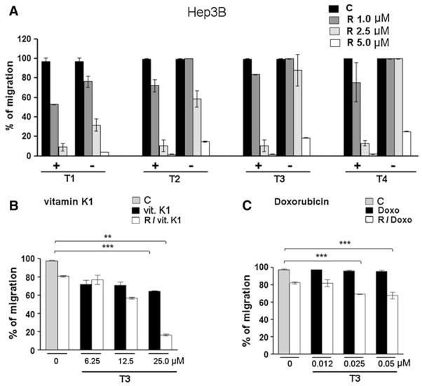
Methods: Cell proliferation, motility, and invasion were analyzed by MTT, wound healing, and invasion assays, respectively, and MAPK pathway protein markers were analyzed by Western blot.

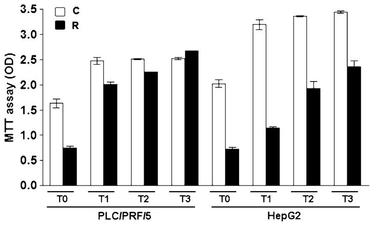
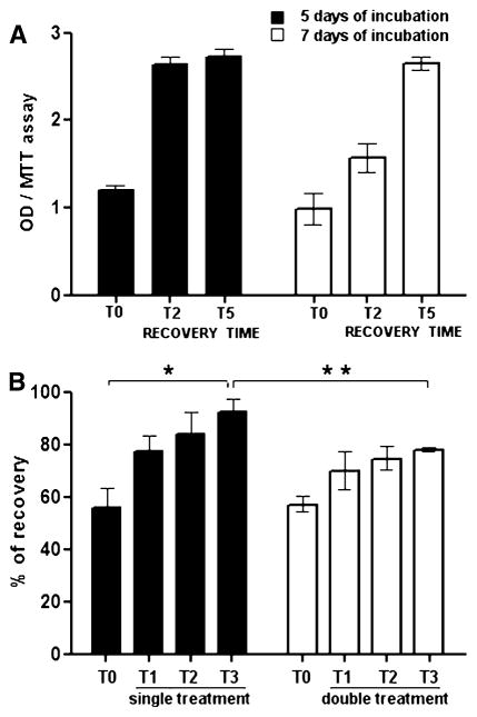
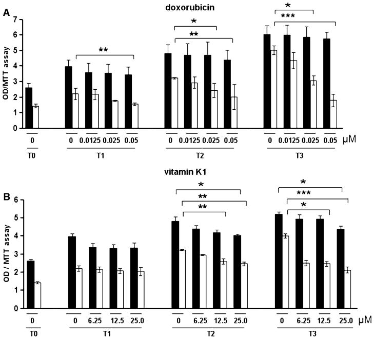
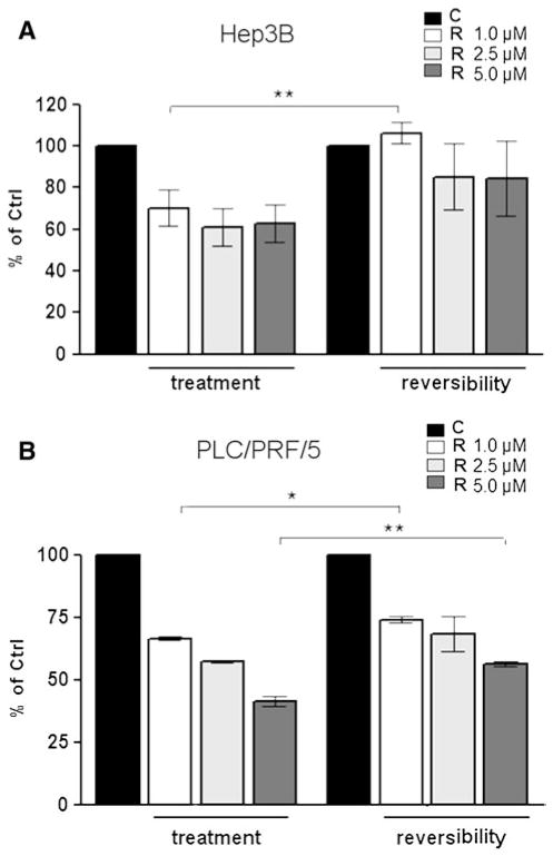
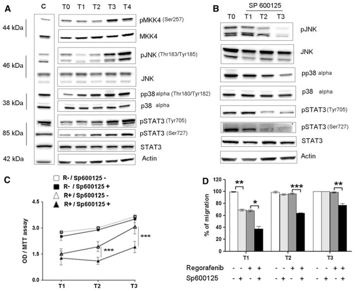
Results: After regorafenib removal, cell growth, migration, and invasion recovered. Repeated drug exposure resulted in changes in cell growth patterns. Recovery could be blocked by sub-growth-inhibitory concentrations of either doxorubicin or vitamin K1. Recovery of growth was associated with increased phospho-JNK, phospho-p38, and phospho-STAT3 levels. The recovery of growth, migration, and signaling were blocked by a JNK inhibitor.

Conclusions: Removal of regorafenib from growth-inhibited cells resulted in a JNK-dependent recovery of growth and migration.


Figures

Fig. 1
Fig. 1
Effects of regorafenib on cell growth recovery in PLC/PRF/5 and HepG2 cell lines. The PLC/PRF/5 and HepG2 cell lines were cultured in regorafenib 5 μM for 72 h. The medium was then removed (T0), and the cells were cultured for the indicated further days (T1–T3) in drug-free medium and analyzed using the MTT assay. The percentage of recovery of the treated cells respect control (vehicle) cells (100 % at T3) was 100 and 60 % for PLC/PRF/5 and HepG2, respectively
Fig. 2
Fig. 2
Recovery of growth after regorafenib-mediated inhibition. a Hep3B cells were cultured in regorafenib 5 μM for 5 or 7 days to inhibit growth. The medium was then removed (T0), and the cells were cultured for the indicated further days in drug-free medium and analyzed using the MTT assay. b Hep3B cells were doubly treated with regorafenib or vehicle and followed after each treatment for growth recovery at different times (T1–T3), using the MTT assay. Values were expressed as percentage of recovery of the treated cells respect control (vehicle) cells (100 % at T3). *P < 0.05, **P < 0.001
Fig. 3
Fig. 3
Effects of doxorubicin and vitamin K1 on cell growth recovery. Hep3B cells were treated with regorafenib 5 μM (white columns) or vehicle (black columns). After medium renewal (T0), the cells cultured with the indicated concentrations of doxorubicin (a) or vitamin K1 (b) were followed for growth recovery at different times (T1–T3) using the MTT assay. *P < 0.05, **P < 0.001, ***P < 0.0001
Fig. 4
Fig. 4
Effects of doxorubicin or vitamin K1 on recovery of cell migration. a Cells treated with different regorafenib concentrations (+) were compared with cells after removal of the same drug concentrations (−) and followed for recovery of migration at different times (T1–T4) after the scratch (T0). (b, c) Cells were treated with regorafenib or vehicle. Medium was then removed (T0) and cells cultured in medium containing the indicated concentrations of vitamin K1 (B) or doxorubicin (C) and followed for recovery of migration. Values were expressed as percentage of migration, 100 % representing the completely closed wound. The symbols minus and plus are two cell groups: cell treated with different concentrations of regorafenib (+) versus cells that, after regorafenib treatment, are cultured in fresh medium without drug (−). The gray bars represent non-drug-treated cells (c). C: vehicle, R: regorafenib. *P < 0.05, **P < 0.001, ***P < 0.0001
Fig. 5
Fig. 5
Recovery of cell invasion after regorafenib treatment. Hep3B (a) and PLC/PRF/5 (b) cells were treated with different concentrations of regorafenib or vehicle. The first data set (called “treatment”) represents the percentage of the invading drug-treated cells compared to drug-untreated control cells, and the second one (called “reversibility”) represents the percentage of the invading recovered cells (after 72 h from drug removal) compared to drug-untreated control cells. C: vehicle, R: regorafenib. *P< 0.05, **P< 0.001
Fig. 6
Fig. 6
Mechanisms of cell growth and migration recovery in Hep3B cells. a Recovery of cells pre-treated with regorafenib 5 μM for 72 h. Phospho-JNK (pJNK) and pp38 levels were increased during reversibility (T1–T4), as were pMKK4 and pSTAT3 (Ser727 and Tyr705) levels, which are upstream and downstream of JNK, respectively. b To test the significance of pJNK changes, SP600125 (JNK inhibitor) 20 μM was used during reversibility. The inhibition of the induced increase in pJNK, pp38, and pSTAT3 (Ser727 and Tyr705) kinase levels (T1–T3) was observed. c, d Cell growth (C) and migration (D) during recovery (T1–T3). R− and R + represent the cells without and with regorafenib treatment (5 μM), respectively. Sp− and Sp + represent cells without or with JNK inhibitor treatment (20 μM), respectively. *P< 0.05, **P< 0.001, ***P< 0.0001

References

    1. Ahmad T, Eisen T. Kinase inhibition with BAY 43-9006 in renal cell carcinoma. Clin Cancer Res. 2004;10:6388S–6392S. - PubMed
    1. Cervello M, Bachvarov D, Lampiasi N, Cusimano A, Azzolina A, McCubrey JA, Montalto G. Molecular mechanisms of sorafenib action in liver cancer cells. Cell Cycle. 2012;11:2843–2855. - PubMed
    1. Wilhelm SM, Carter C, Tang L, Wilkie D, McNabola A, Rong H, Chen C, Zhang X, Vincent P, McHugh M, Cao Y, Shujath J, Gawlak S, Eveleigh D, Rowley B, Liu L, Adnane L, Lynch M, Auclair D, Taylor I, Gedrich R, Voznesensky A, Riedl B, Post LE, Bollag G, Trail PA. BAY 43-9006 exhibits broad spectrum oral antitumor activity and targets the RAF/MEK/ERK pathway and receptor tyrosine kinases involved in tumor progression and angiogenesis. Cancer Res. 2004;64:7099–7109. - PubMed
    1. Liu L, Cao Y, Chen C, Zhang X, McNabola A, Wilkie D, Wilhelm S, Lynch M, Carter C. Sorafenib blocks the RAF/MEK/ERK pathway, inhibits tumor angiogenesis, and induces tumor cell apoptosis in hepatocellular carcinoma model PLC/PRF/5. Cancer Res. 2006;66:11851–11858. - PubMed
    1. Llovet JM, Ricci S, Mazzaferro V, Hilgard P, Gane E, Blanc JF, de Oliveira AC, Santoro A, Raoul JL, Forner A, Schwartz M, Porta C, Zeuzem S, Bolondi L, Greten TF, Galle PR, Seitz JF, Borbath I, Häussinger D, Giannaris T, Shan M, Moscovici M, Voliotis D, Bruix J SHARP Investigators Study Group . Sorafenib in advanced hepatocellular carcinoma. N Engl J Med. 2008;359:378–390. - PubMed

Publication types

MeSH terms