. 2014 Feb;59(2):630-42.
doi: 10.1002/hep.26697. Epub 2013 Dec 23.

Chemokine receptor CCR6-dependent accumulation of γδ T cells in injured liver restricts hepatic inflammation and fibrosis

Affiliations

Chemokine receptor CCR6-dependent accumulation of γδ T cells in injured liver restricts hepatic inflammation and fibrosis

Linda Hammerich et al. Hepatology. 2014 Feb.

Abstract

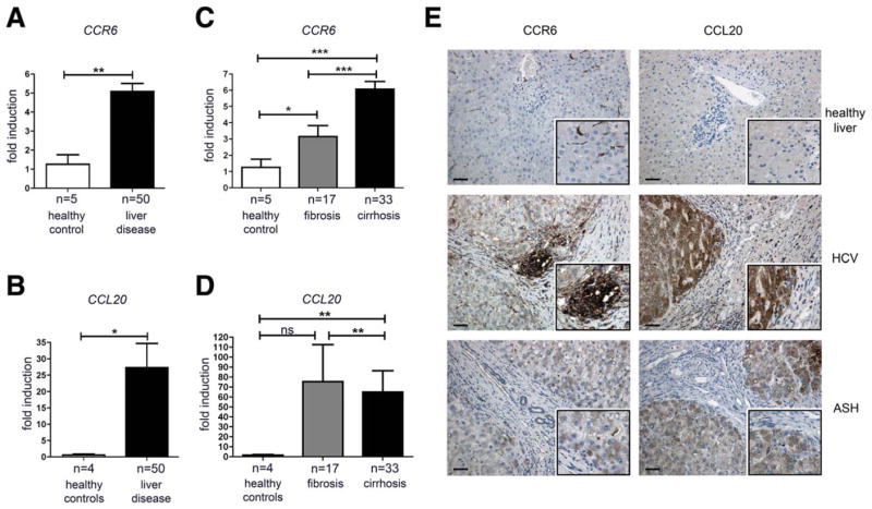
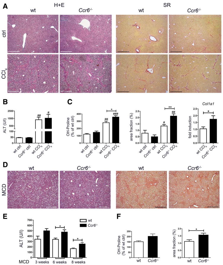
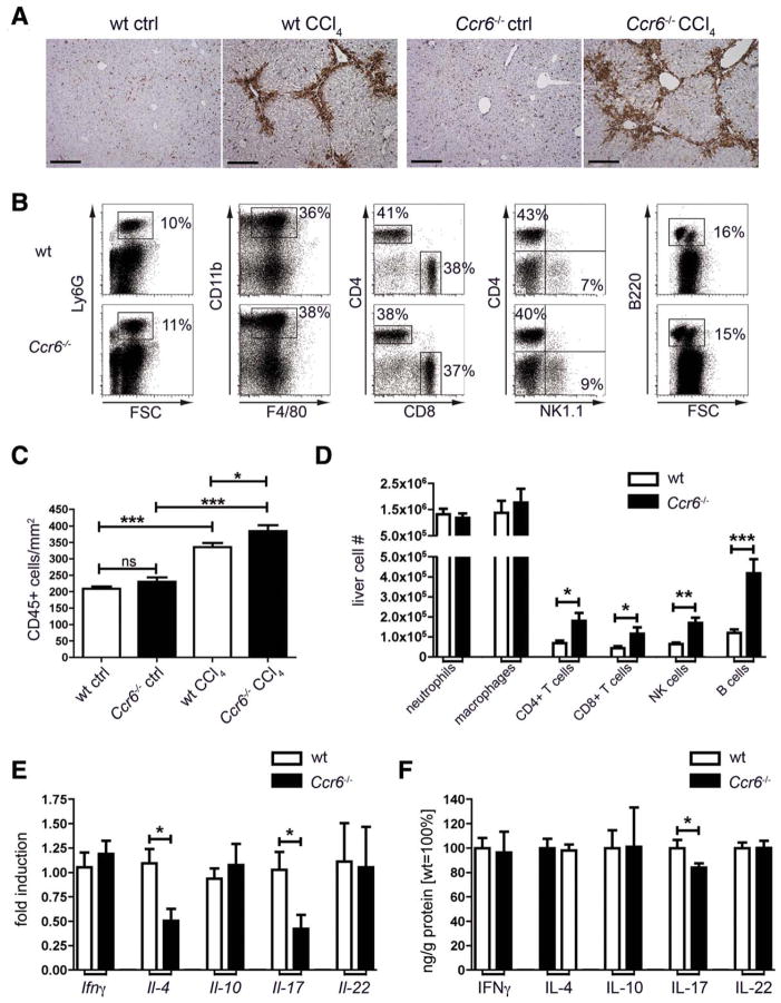
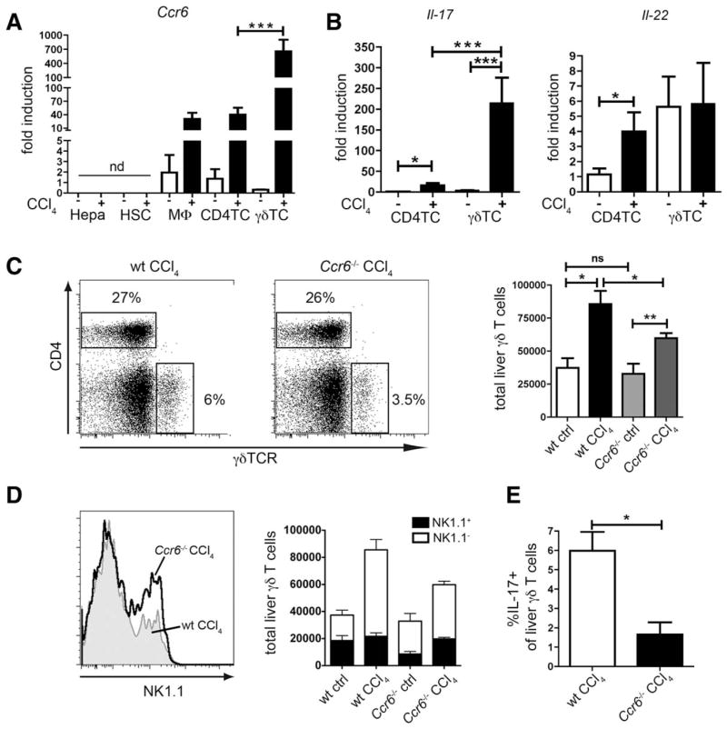
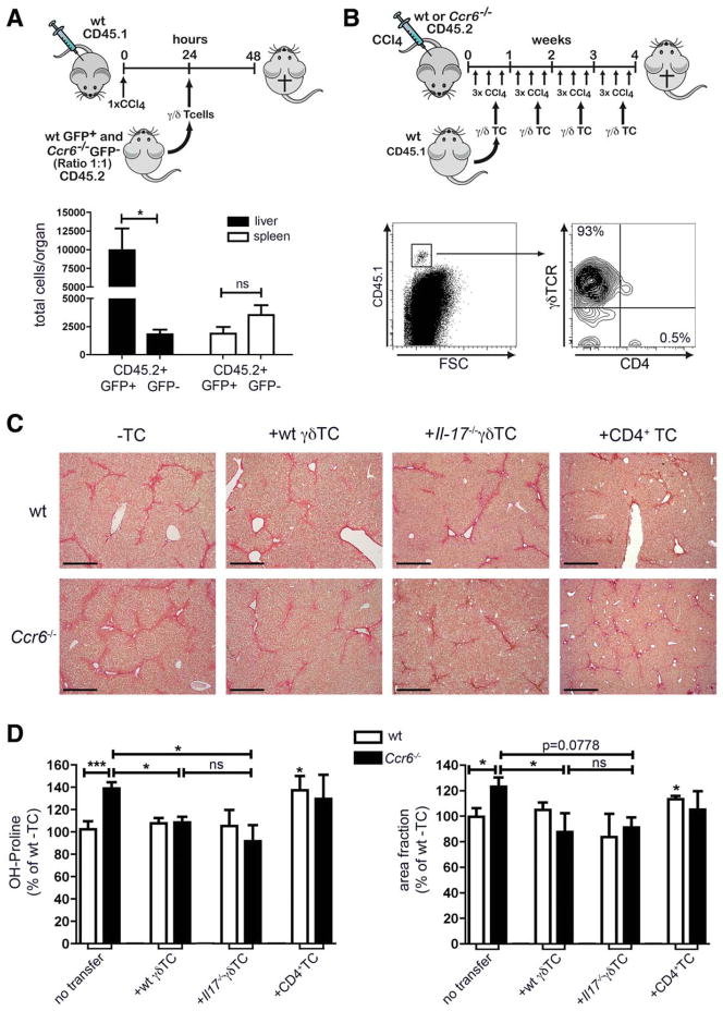
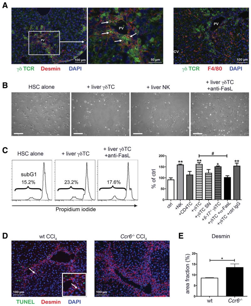
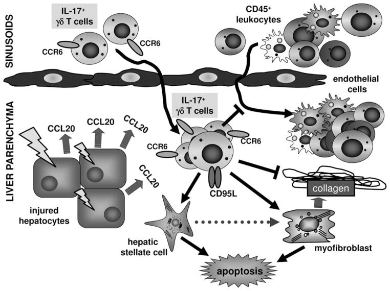
Chronic liver injury promotes hepatic inflammation, representing a prerequisite for organ fibrosis. We hypothesized a contribution of chemokine receptor CCR6 and its ligand, CCL20, which may regulate migration of T-helper (Th)17, regulatory, and gamma-delta (γδ) T cells. CCR6 and CCL20 expression was intrahepatically up-regulated in patients with chronic liver diseases (n = 50), compared to control liver (n = 5). Immunohistochemistry revealed the periportal accumulation of CCR6(+) mononuclear cells and CCL20 induction by hepatic parenchymal cells in liver disease patients. Similarly, in murine livers, CCR6 was expressed by macrophages, CD4 and γδ T-cells, and up-regulated in fibrosis, whereas primary hepatocytes induced CCL20 upon experimental injury. In two murine models of chronic liver injury (CCl4 and methionine-choline-deficient diet), Ccr6(-/-) mice developed more severe fibrosis with strongly enhanced hepatic immune cell infiltration, compared to wild-type (WT) mice. Although CCR6 did not affect hepatic Th-cell subtype composition, CCR6 was explicitly required by the subset of interleukin (IL)-17- and IL-22-expressing γδ T cells for accumulation in injured liver. The adoptive transfer of WT γδ, but not CD4 T cells, into Ccr6(-/-) mice reduced hepatic inflammation and fibrosis in chronic injury to WT level. The anti-inflammatory function of hepatic γδ T cells was independent of IL-17, as evidenced by transfer of Il-17(-/-) cells. Instead, hepatic γδ T cells colocalized with hepatic stellate cells (HSCs) in vivo and promoted apoptosis of primary murine HSCs in a cell-cell contact-dependent manner, involving Fas-ligand (CD95L). Consistent with γδ T-cell-induced HSC apoptosis, activated myofibroblasts were more frequent in fibrotic livers of Ccr6(-/-) than in WT mice.

Conclusion: γδ T cells are recruited to the liver by CCR6 upon chronic injury and protect the liver from excessive inflammation and fibrosis by inhibiting HSCs.


Conflict of interest statement

Potential conflict of interest: Nothing to report.

Figures

Fig. 1
Fig. 1
CCR6 and CCL20 are up-regulated in human CLD. (A–D) Liver samples of patients with CLD and control tissue were analyzed for CCR6 and CCL20 expression levels by quantitative real-time polymerase chain reaction, normalized to β-actin. (E) Human liver sections were stained for CCR6 and CCL20; representative pictures from n = 3 individual patients per group are shown. Inserts display higher magnifications of positively stained areas. Scale bar: 100 μm. HCV, chronic hepatitis C virus infection, ASH, alcoholic steatohepatitis. *P < 0.05; **P < 0.01; ***P < 0.001. Data are shown as mean ± standard error of the mean.
Fig. 2
Fig. 2
CCR6 deficiency promotes hepatic fibrosis in mice. (A–C) WT and Ccr6−/− mice were challenged with CCl4 thrice-weekly for 4 weeks or left untreated. (A) Representative hematoxylin and eosin (H+E) and Sirius Red (SR) stainings of WT and Ccr6−/− mice. (B) Alanine aminotransferase (ALT) serum activities in WT and Ccr6−/− mice. (C) Hydroxyproline (OH-Proline) measurement, quantification of matrix deposition from Sirius Red stainings (derived from stainings as shown in A) and expression of Collagen1a1 (Col1a1) in livers of WT and Ccr6−/− mice. (D–F) WT and Ccr6−/− mice were fed with MCD diet for 8 weeks to induce steatohepatitis. (D) Representative H+E and Sirius Red stainings. (E) Serum ALT levels at indicated time points. (F) Hydroxyproline measurement and quantification of matrix deposition from livers of WT and Ccr6−/− mice. */#P < 0.05; **/##P < 0.01; ***/###P < 0.001. #Compared to control conditions. Scale bars: 500 μm. Data are derived from n = 7–10 mice per group and shown as mean ± standard error of the mean. To accurately compare results for fibrosis from different experiments, data were normalized to the respective WT control liver (=100%) from the same experiment.
Fig. 3
Fig. 3
Leukocyte infiltration and Th subset composition in injured livers of WT and CCR6-deficient mice. Wt and Ccr6−/− mice were treated thrice-weekly with CCl4 for 4 weeks or left untreated. (A) IHC for the pan-leukocyte marker CD45 in livers of WT and Ccr6−/− mice. Scale bar: 200 μm. (B) Liver leukocytes (CD45+, Hoechst-33258 negative) were subdivided into neutrophils (Ly6G+), macrophages (CD11b+F4/80+), T cells (CD3+CD4+, CD3+CD8+), NK cells (CD3NK1.1+), and B cells (B220+). Representative FACS plots from livers of CCl4-treated mice. (C) Quantification of (A). (D) Absolute numbers of intrahepatic leukocyte subpopulations shown in (B). (E) Expression of signature cytokines for CD4 Th cell subtypes was measured by quantitative polymerase chain reaction from total liver, namely, Ifnγ (Th1), Il-4 (Th2), Il-10 (Treg), and Il-17 and Il-22 (Th17). (F) Concentrations of cytokines were measured by enzyme-linked immunosorbent assay from liver and normalized to total protein content in liver extracts. Because of different absolute cytokine concentrations, values are displayed as % of fibrotic WT liver. *P < 0.05; **P < 0.01; ***P < 0.001. Data are derived from n = 6–10 mice per group and shown as mean ± standard error of the mean.
Fig. 4
Fig. 4
Intrahepatic accumulation of IL-17 producing γδ T cells is impaired in Ccr6−/− mice upon chronic injury. (A) Hepatocytes (Hepa), HSC, macrophages (MΦ), CD4+ T cells (CD4TC), and γδ T cells (γδ TC) were isolated by FACS sorting from livers of WT mice treated with CCl4 or control mice. Ccr6 expression was determined by quantitative polymerase chain reaction. (B) CD4+ T cells and γδ T cells were FACS sorted from WT livers. Il-17 and Il-22 expression by quantitative polymerase chain reaction. (C) Absolute numbers of γδ T cells (CD3+CD4γδTCR+) in livers of WT or Ccr6−/− mice treated with CCl4 or control mice. Representative FACS plots from livers of CCl4-treated mice. (D and E) Same as (C) with additional staining for NK1.1 and IL-17 after restimulation with phorbol 12-myristate 13-acetate PMA/ionomycin to discriminate γδ T-cell subtypes. (D) Representative histograms of NK1.1 expression by hepatic γδ T cells (CCl4-treated mice, gated on CD3+CD4γδTCR+) and quantification of NK1.1+ and NK1.1 γδ T cells. (E) Quantification of IL-17+ γδ T cells in the liver (CCl4-treated mice, IL-17 expression determined by intracellular FACS staining). *P < 0.05; **P < 0.01; ***P < 0.001. Data are derived from n = 6–10 mice per group and shown as mean ± standard error of the mean.
Fig. 5
Fig. 5
Reconstitution of Ccr6−/− mice with γδ T cells, but not with CD4+ T cells, reduces hepatic fibrosis. (A) Splenic γδ T cells were isolated from actin-eGFP or Ccr6−/− mice, mixed in a 1:1 ratio and injected into CD45.1+ B6-mice that had been treated with CCl4 once (24 hours before T-cell injection). Twenty-four hours after T-cell injection absolute numbers of CD45.2+GFP+ WT and CD45.2+GFP Ccr6−/− γδ T cells in liver and spleen were determined. (B–D) WT and Ccr6−/− mice were treated with CCl4 thrice-weekly over 4 weeks. Simultaneously, splenic CD4+ or γδ T cells were isolated from CD45.1+ mice and injected IV once per week. Mice received either wt γδ T cells (100,000–150,000 cells), Il-17−/− γδ T cells (100,000–150,000 cells), or WT CD4+ T cells (1,000,000–1,500,000 cells) during CCl4 treatment. (B) Leukocytes were isolated from liver and stained for CD45.1, CD4, and γδ TCR to identify transferred cells (representative plot shown). (C) Sirius Red stainings of liver paraffin sections. Scale bar: 500 μm. (D) Measurement of hepatic hydroxyproline and quantification of ECM deposition. *P < 0.05. Data are derived from n = 6 mice per group and shown as mean ± standard error of the mean. To accurately compare results for fibrosis from different experiments, data were normalized to the respective WT liver without cell transfer (=100%) from the same experiment.
Fig. 6
Fig. 6
γδ T cells induce HSC apoptosis. (A) WT mice were treated thrice-weekly with CCl4 for 4 weeks. Liver sections were stained for γδ TCR to identify γδ T cells (green) and desmin to identify HSC (red, left and middle panel) or F4/80 to identify macrophages (red, right panel). Nuclei were counterstained with DAPI (blue). PV, portal vein; CV, central vein. (B and C) Hepatic CD4+ T cells, γδ T cells, and NK cells were isolated by FACS sorting from WT mice 48 hours after a single CCl4 injection. Either isolated cells or supernatant from overnight cultures were incubated with primary HSCs, isolated from WT mice, for 48 hours. (B) Representative pictures of cocultures. Scale bar: 200 μm. (C) HSCs were fixed, permeabilized, and stained with PI for FACS analysis. Representative histograms with sub-G1 cells are shown. Amounts of apoptotic cells are shown as percent of control. (D) WT and Ccr6−/− mice were treated thrice-weekly with CCl4 for 4 weeks. Liver sections were stained for desmin to identify HSCs (red) and TUNEL to identify apoptotic cells (green). Nuclei were counterstained with DAPI (blue). Arrow indicates a representative apoptotic HSC in liver of CCl4-treated WT mice. (E) Quantification of desmin-positive areas in pictures shown in (D). */#P < 0.05; **P < 0.01; ***P < 0.001. *Compared to control. Data are expressed as mean ± standard error of the mean from three independent experiments. DAPI, 4′,6-diamidino-2-phenylindole.
Fig. 7
Fig. 7
Functional role of CCL20/CCR6 and γδ T cells in liver inflammation and fibrosis. Schematic depiction summarizing results from in vitro and in vivo experiments. Upon liver injury, hepatocytes and (especially in cholestatic diseases) injured large bile ducts induce expression of the chemokine CCL20 thereby attracting CCR6+ IL-17+ γδ T cells into injured liver. These γδ T cells reduce hepatic inflammation and fibrosis in chronic injury, independent of its signature cytokine IL-17. One important mechanism appears to be that hepatic γδ T cells promote apoptosis of HSCs and myofibroblasts in a cell-cell contact-dependent manner, involving FasL (CD95L).

Similar articles

Cited by

References

    1. Bataller R, Brenner DA. Liver fibrosis. J Clin Invest. 2005;115:209–218. - PMC - PubMed
    1. Zimmermann HW, Tacke F. Modification of chemokine pathways and immune cell infiltration as a novel therapeutic approach in liver inflammation and fibrosis. Inflamm Allergy Drug Targets. 2011;10:509–536. - PubMed
    1. Shields PL, Morland CM, Salmon M, Qin S, Hubscher SG, Adams DH. Chemokine and chemokine receptor interactions provide a mechanism for selective T cell recruitment to specific liver compartments within hepatitis C-infected liver. J Immunol. 1999;163:6236–6243. - PubMed
    1. Oo YH, Banz V. CXCR3 dependent recruitment and CCR6 mediated positioning of Th-17 cells in the inflamed liver. J Hepatol. 2012;57:1044–1051. - PMC - PubMed
    1. Bonacchi A, Petrai I, Defranco RM, Lazzeri E, Annunziato F, Efsen E, et al. The chemokine CCL21 modulates lymphocyte recruitment and fibrosis in chronic hepatitis C. Gastroenterology. 2003;125:1060–1076. - PubMed

Publication types

MeSH terms