. 2013 Oct 1;73(19):6080-93.
doi: 10.1158/0008-5472.CAN-13-0926. Epub 2013 Aug 19.

Obesity promotes breast cancer by CCL2-mediated macrophage recruitment and angiogenesis

Affiliations

Obesity promotes breast cancer by CCL2-mediated macrophage recruitment and angiogenesis

Lisa M Arendt et al. Cancer Res. .

Abstract

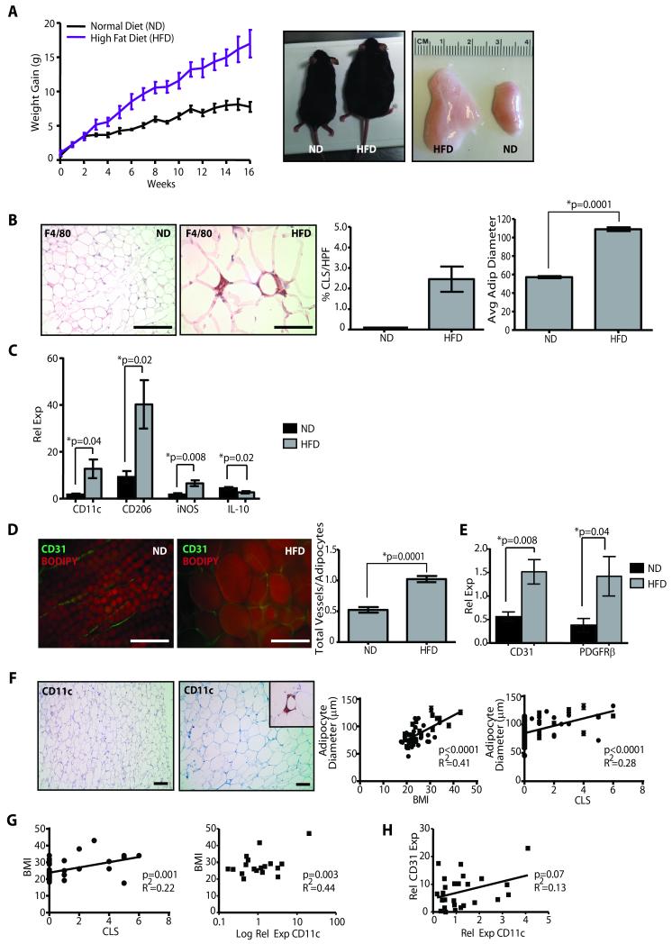
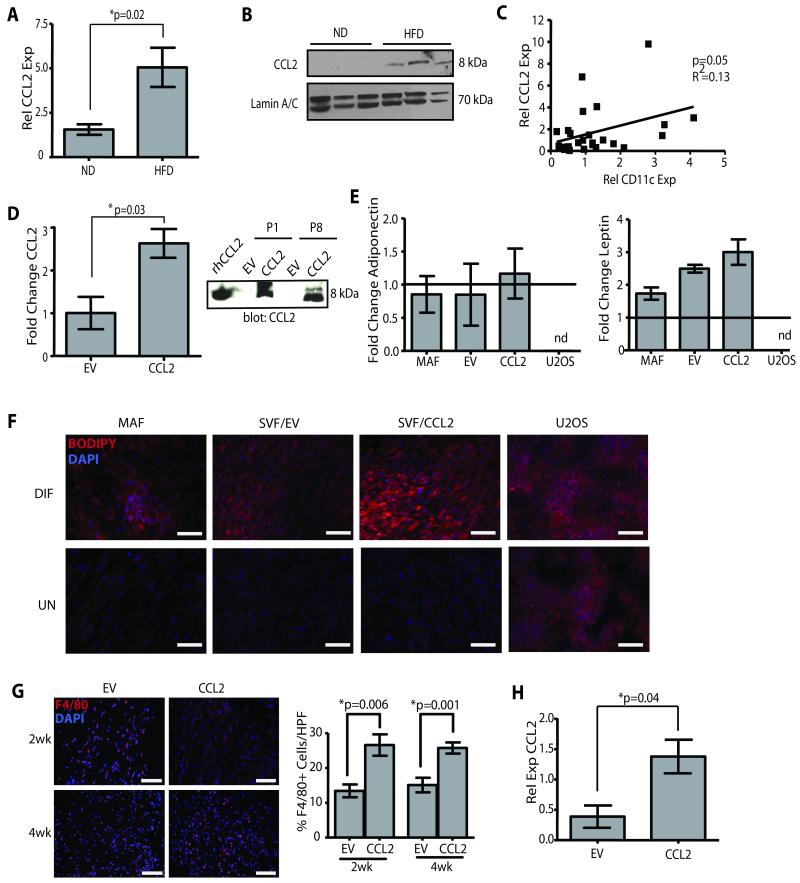
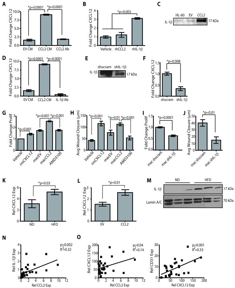
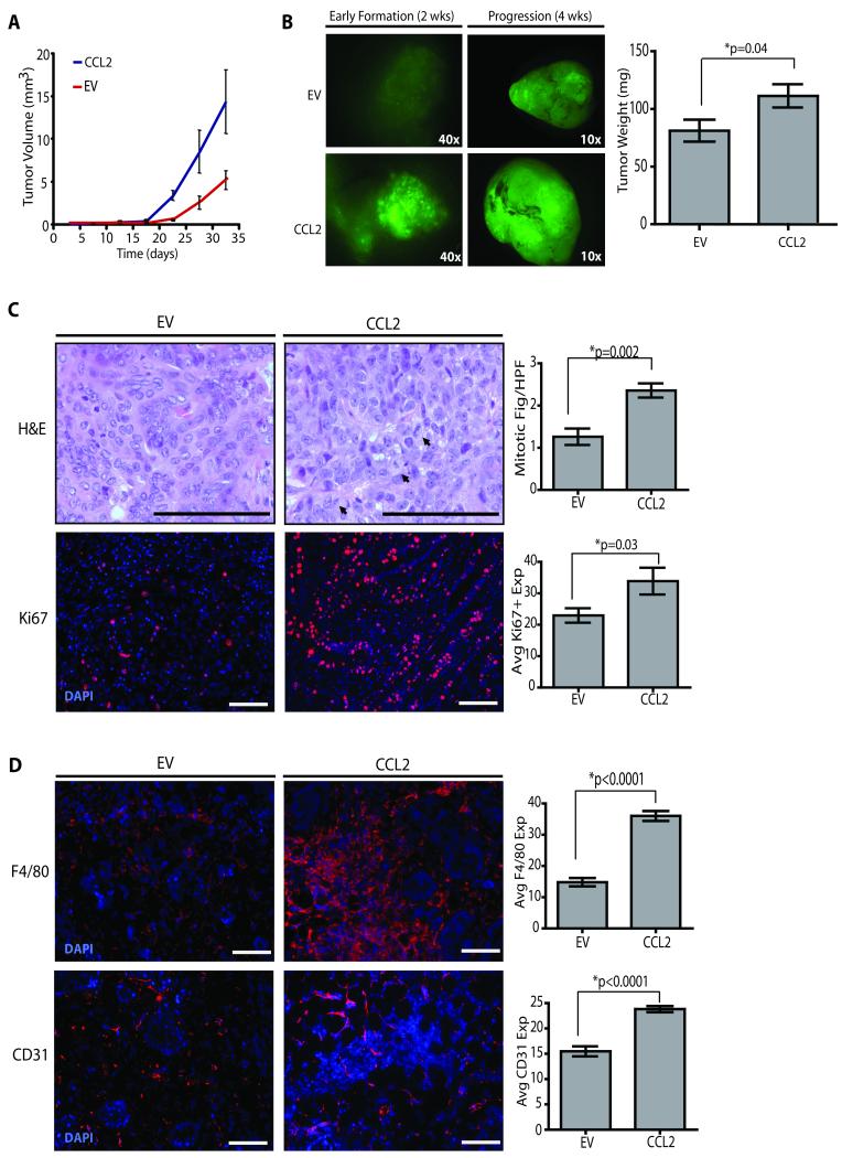
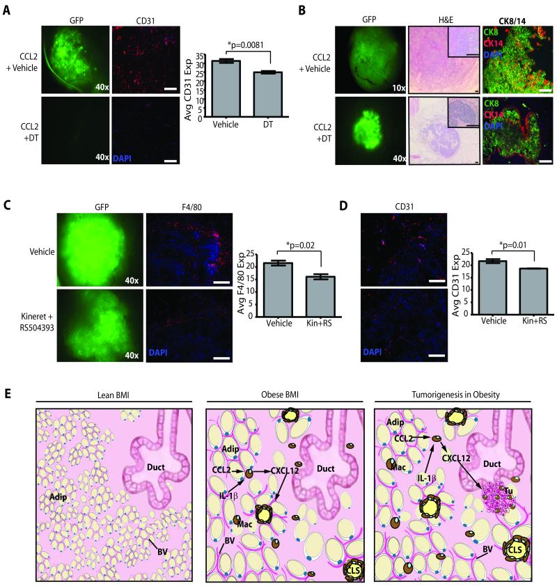
Obesity is one of the most important preventable causes of cancer and the most significant risk factor for breast cancer in postmenopausal women. Compared with lean women, obese women are more likely to be diagnosed with a larger, higher grade tumor, an increased incidence of lymph node metastases, and elevated risk of distant recurrence. However, the mechanisms connecting obesity to the pathogenesis of breast cancer are poorly defined. Here, we show that during obesity, adipocytes within human and mouse breast tissues recruit and activate macrophages through a previously uncharacterized CCL2/IL-1β/CXCL12 signaling pathway. Activated macrophages in turn promote stromal vascularization and angiogenesis even before the formation of cancer. Recapitulating these changes using a novel humanized breast cancer model was sufficient to promote angiogenesis and prime the microenvironment prior to neoplastic transformation for accelerated breast oncogenesis. These findings provide a mechanistic role for adipocytes and macrophages before carcinogenesis that may be critical for prevention and treatment of obesity-related cancer.


Figures

Figure 1
Figure 1. Mammary stroma recruits inflammatory macrophages and demonstrates increased vascularity under conditions of obesity
(A) Female C57Bl/6 mice fed a high fat diet (HFD) gained significantly more weight, resulting in increased body weight (p=0.0006, n=10 mice/group) and mammary fat pad depot size (p=0.002), compared to those fed a normal chow diet (ND). (B) In obese mice, mammary fat depots demonstrated F4/80+ crown-like structures (CLS), which were not detectable in depots of control mice, as well as significantly increased adipocyte diameters. (C) Transcripts for M1-like (CD11c, iNOS) and M2-like (CD206) macrophage markers were elevated in mammary glands from HFD mice (qPCR, n=6 mice/group). (D) Mammary glands from obese mice demonstrated a significantly increased vessel to adipocyte ratio (n=5 mice/group). (E) Transcripts for CD31 and PDGFRβ were significantly elevated in glands from HFD mice (qPCR, n=6/group). (F) In breast tissue isolated from reduction mammoplasty surgeries, increasing adipocyte diameter was correlated with increased body mass index (BMI; n=46) as well as the formation of CD11c+ CLS (n=76). (G) Increased BMI was correlated with the formation of CD11c+ CLS (n=46), as well as increased relative expression of CD11c transcripts from reduction mammoplasty tissues (n=18). (H) Elevated CD11c expression was associated with increased CD31 expression (n=28). qPCR was performed on RNA isolated from reduction mammoplasty tissue. Original magnification (B, D) 200x, (F) 100x; bar=100μm.
Figure 2
Figure 2. Overexpression of CCL2 in human adipose stroma cells results in obesity-like conditions
(A) CCL2 transcripts were significantly increased in mammary glands of C57Bl/6 mice fed HFD diet compared to those from ND mice (qPCR, n=6 mice/group). (B) CCL2 protein was elevated in glands from obese mice. Lamin A/C was used as a loading control. (C) Increased expression of CCL2 and CD11c were significantly correlated in mammary reduction tissue (qPCR, n=28 samples). (D) SVF/CCL2 cells expressed significantly increased levels of CCL2 mRNA and protein in conditioned media compared with SVF/EV cells (n=3 experiments). Recombinant human CCL2 (rhCCL2) was used as a positive control. (E) Mature adipocyte fraction (MAF), SVF/EV, SVF/CCL2, and U2OS cells were treated with adipogenic media (DIF) or vehicle (UN). Following differentiation, changes in expression levels of adiponectin and leptin were similar to the MAF but were not detected (nd) in U2OS cells. qPCR was performed on RNA isolated from 3 experiments, and data represented as fold change following differentiation compared with vehicle-treated cells. (F) Stromal cells and U2OS cells were exposed to adipogenic media or vehicle; lipid was stained with BODIPY 558/568 and counterstained with DAPI. (G) Mammary glands from NOD/SCID mice humanized with SVF/CCL2 cells demonstrated increased F4/80+ cells compared with glands humanized with SVF/EV cells (n=3 mice/group). (H) After 4 weeks, CCL2 transcripts were significantly increased in glands from NOD/SCID mice humanized with SVF/CCL2 compared with glands humanized with SVF/EV cells. Original magnification (F, G) 200x; bar=100μm.
Figure 3
Figure 3. Macrophages promote angiogenesis in response to obesity-like stromal cells
(A, B) Mammary glands from NOD/SCID mice humanized with SVF/CCL2 cells demonstrated significantly increased CD31+ cells after 4 weeks compared with glands humanized with SVF/EV cells as quantified by immunofluorescence (A) and flow cytometry (B). Glands from 3 mice were utilized for each time point. (C) Matrigel plugs were implanted under the flank of Mac-SCID mice. Those containing SVF/CCL2 cells recruited significantly increased numbers of F4/80+ macrophages and CD31+ endothelial cells than plugs containing SVF/EV cells. In mice treated with diphtheria toxin (DT), both F4/80+ macrophages and CD31+ endothelial cells were significantly decreased in plugs containing either SVF/CCL2 or SVF/EV cells compared with plugs from vehicle treated mice. (D) At 2 weeks following humanization, DT treatment decreased both CD11b+ and CD31+ cells compared with glands from vehicle-treated mice. Four weeks following humanization and DT administration, no significant changes in CD11b+ cells were observed, and only DT-treated SVF/CCL2 glands demonstrated significantly reduced CD31+ cells compared with those from vehicle-treated mice. Mice received DT or vehicle at 24 hours and 48 hours following humanization. Glands were collected at 2 or 4 weeks following DT administration (n=5 mice/group). Two Matrigel plugs were injected subcutaneously in each mouse. Data denoted as a fold change from vehicle treated mice in each condition. Original magnification (A) 200x; bar=100μm.
Figure 4
Figure 4. Macrophage derived CXCL12 induces angiogenesis
(A) SVF/CCL2 conditioned media (CM) significantly increased CXCL12 expression in HL-60 macrophages compared with SVF/EV CM or SVF/CCL2 CM + blocking antibody for CCL2 (CCL2 Ab). (B) Recombinant human IL-1β (rhIL-1β) increased CXCL12 expression in HL-60 macrophages. qPCR was performed on RNA isolated from 3 experiments. (C) SVF/CCL2 CM contained increased IL-1β protein compared to SVF/EV CM. CM from HL-60 macrophages treated with LPS was used as a positive control. (D) SVF/CCL2 CM+IL-1β blocking Ab significantly reduced CXCL12 expression in HL-60 macrophages compared to SVF/CCL2 CM. (E) CM from SVF/CCL2 shIL-1β (shIL-1β) cells demonstrated decreased IL-1β protein compared to CM from SVF/CCL2 shscrambled control cells (shscram). (F) SVF/CCL2 shIL-1β CM significantly decreased CXCL12 expression in HL-60 macrophages compared with SVF/CCL2 shscramble CM. (G, H) Human microvascular endothelial cells (HMVEC) proliferated (G) and migrated (H) in response to recombinant mouse CXCL12 (rmCXCL12), as well as in response to CM from HL-60 macrophages pre-treated with CM from SVF/CCL2 cells (macCCL2) compared to those treated with SVF/EV CM (macEV). AMD3100+macCCL2 CM significantly decreased HMVEC proliferation and migration. Proliferation data are represented as a fold change of vehicle-treated cells, and 3 experiments were performed in triplicate. (I, J) HMVEC demonstrated decreased proliferation (I) and migration (J) in response to CM from HL-60 macrophages pre-treated with CM from SVF/CCL2 shIL-1β cells (mac shIL-1β) compared to CM from SVF/CCL2 shscrambled cells (mac shscram). Proliferation data are represented as a fold change of vehicle-treated cells, and 3 experiments were performed in triplicate. (K) Glands from C57Bl/6 HFD mice demonstrated elevated CXCL12 expression compared to glands from ND mice (n=6 mice/group). (L) SVF/CCL2 humanized glands from NOD/SCID mice demonstrated significantly increased CXCL12 expression compared to SVF/EV humanized glands (n=6 mice/group). (M) IL-1β protein was increased in C57Bl/6 HFD mammary glands compared to ND glands. (N, O) Reduction mammoplasty tissue demonstrated a significant correlation between CCL2 and IL-1β expression levels (N), as well as significant correlation between expression of CXCL12 and both CCL2 and CD31 (O) qPCR was performed on RNA isolated from 28 reduction mammoplasty samples.
Figure 5
Figure 5. Obesity-like stromal cells accelerate tumorigenesis
(A) Tumors from glands from NOD/SCID mice humanized with SVF/CCL2 cells developed earlier than those from SVF/EV humanized glands. Human mammary epithelial cells (HMEC) were isolated from 3 reduction mammoplasty tissue samples, transduced with lentiviruses encoding SV40er and KrasG12V, and transplanted into humanized glands co-mixed with either SVF/CCL2 or SVF/EV cells. (B) Tumors from SVF/CCL2 humanized glands were significantly larger in size and weight at four weeks after transplantation. N=6 tumors/group from 3 experiments with separate patient tissue samples (Table S1). (C) Tumors arising in SVF/CCL2 glands demonstrated significantly increased numbers of mitotic figures and Ki67 expression compared with those from SVF/EV glands. Mitotic figures and Ki67+ cells were quantified from 3 images from 12 tumors/group. Statistical differences were detected by t-test. (D) At 2 weeks following transplantation of transduced HMECs, glands humanized with SVF/CCL2 cells demonstrated significantly increased F4/80+ macrophage recruitment and increased CD31+ cells (n=6 tumors/group). Original magnification (C) 600x, 200x; (D) 200x; bar=100μm.
Figure 6
Figure 6. Stromal changes associated with obesity accelerate tumorigenesis
(A) Two weeks following transplantation of virally transduced human mammary epithelial cells (HMECs), diphtheria toxin (DT)-treated mice demonstrated reduced transplant growth and significantly reduced CD31+ cells compared with vehicle-treated mice (n=6 tumors/group). (B) At end stage, glands of vehicle-treated Mac-SCID mice developed large tumors with extensive stromalization and invasive borders, with cytokeratin (CK) 8+ and CK14+ cells randomly distributed in the tumor paremchyma. Diphtheria toxin (DT)-treated mice developed small, well-circumscribed growths with CK8+ cells surrounded by a ring of CK14+ cells. (C, D) Two weeks following transplant of virally transformed HMECs, glands from mice treated with Kineret and RS504393 (n=10 transplants/group) demonstrated significantly reduced F4/80+ macrophage infiltration (C), as well as significantly reduced CD31 expression (D). (E) Obese adipose tissue (Adip) surrounding epithelial cells of the mammary gland (Duct) secretes CCL2, leading to the recruitment of inflammatory macrophages (Mac) to form crown-like structures (CLS). In response to CCL2 and IL-1β expression, macrophages secrete CXCL12, which acts on blood vessels (BV) to enhance angiogenesis. Obese adipose tissue is primed for early angiogenesis and inflammatory signaling which promotes aggressive breast malignancies (Tu). Original magnification: (A, C, D) 200x, (B) middle panel 40x, inset 200x, end panel 200x; bar=100μm.

References

    1. Calle EE, Kaaks R. Overweight, obesity and cancer: epidemiological evidence and proposed mechanisms. Nat Rev Cancer. 2004;4:579–91. - PubMed
    1. Calle EE, Rodriguez C, Walker-Thurmond K, Thun MJ. Overweight, obesity, and mortality from cancer in a prospectively studied cohort of U.S. adults. N Engl J Med. 2003;348:1625–38. - PubMed
    1. Carmichael AR. Obesity as a risk factor for development and poor prognosis of breast cancer. BJOG. 2006;113:1160–6. - PubMed
    1. Brown KA, Simpson ER. Obesity and breast cancer: mechanisms and therapeutic implications. Front Biosci (Elite Ed) 2012;4:2515–24. - PubMed
    1. Morris PG, Hudis CA, Giri D, Morrow M, Falcone DJ, Zhou XK, et al. Inflammation and increased aromatase expression occur in the breast tissue of obese women with breast cancer. Cancer Prev Res (Phila) 2011;4:1021–9. - PMC - PubMed

MeSH terms