. 2013 Sep 3;110(36):E3408-16.
doi: 10.1073/pnas.1300600110. Epub 2013 Aug 19.

Inherited mutations in the helicase RTEL1 cause telomere dysfunction and Hoyeraal-Hreidarsson syndrome

Affiliations

Inherited mutations in the helicase RTEL1 cause telomere dysfunction and Hoyeraal-Hreidarsson syndrome

Zhong Deng et al. Proc Natl Acad Sci U S A. .

Abstract

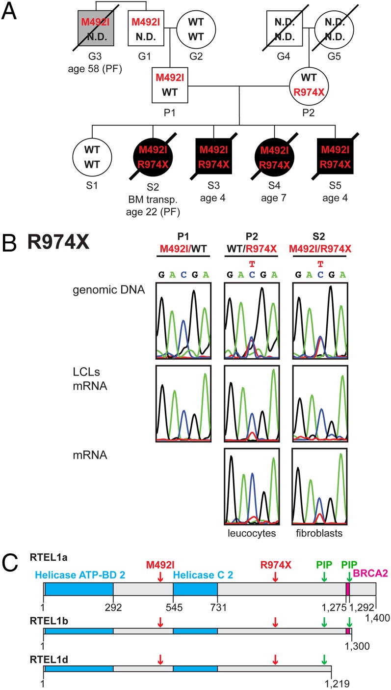
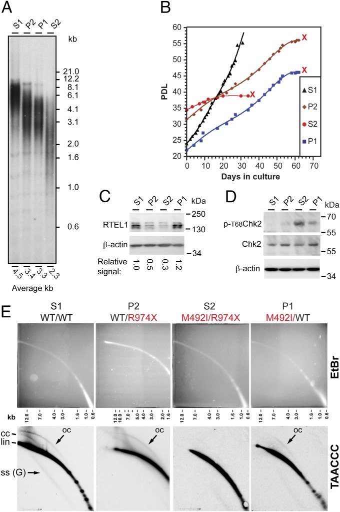
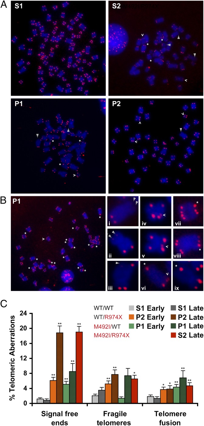
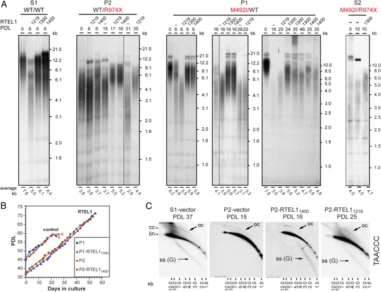
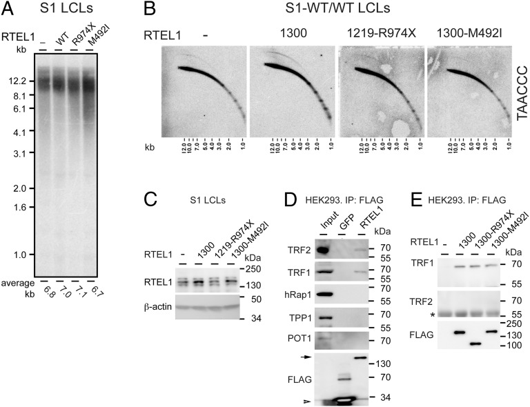
Telomeres repress the DNA damage response at the natural chromosome ends to prevent cell-cycle arrest and maintain genome stability. Telomeres are elongated by telomerase in a tightly regulated manner to ensure a sufficient number of cell divisions throughout life, yet prevent unlimited cell division and cancer development. Hoyeraal-Hreidarsson syndrome (HHS) is characterized by accelerated telomere shortening and a broad range of pathologies, including bone marrow failure, immunodeficiency, and developmental defects. HHS-causing mutations have previously been found in telomerase and the shelterin component telomeric repeat binding factor 1 (TRF1)-interacting nuclear factor 2 (TIN2). We identified by whole-genome exome sequencing compound heterozygous mutations in four siblings affected with HHS, in the gene encoding the regulator of telomere elongation helicase 1 (RTEL1). Rtel1 was identified in mouse by its genetic association with telomere length. However, its mechanism of action and whether it regulates telomere length in human remained unknown. Lymphoblastoid cell lines obtained from a patient and from the healthy parents carrying heterozygous RTEL1 mutations displayed telomere shortening, fragility and fusion, and growth defects in culture. Ectopic expression of WT RTEL1 suppressed the telomere shortening and growth defect, confirming the causal role of the RTEL1 mutations in HHS and demonstrating the essential function of human RTEL1 in telomere protection and elongation. Finally, we show that human RTEL1 interacts with the shelterin protein TRF1, providing a potential recruitment mechanism of RTEL1 to telomeres.

Keywords: aging; dyskeratosis congenita; genomic instability; telomeropathies.


Conflict of interest statement

The authors declare no conflict of interest.

Figures

Fig. 1.
Fig. 1.
Compound heterozygous RTEL1 mutations were associated with HHS. (A) Genealogical tree of the family. Open circles and squares represent unaffected females and males, respectively. Black circles and squares represent affected females and males. A gray square indicates a family member who died from pulmonary fibrosis. Tilted lines indicate mortality, and the ages of mortality are indicated underneath. Patient S2 underwent bone marrow transplantation (BM transp.) but passed away 5 y later from pulmonary fibrosis. (B) PCR amplification and sequencing of exon 30 from genomic DNA validated the presence of the heterozygous R974X mutation in S2 and P2, but not P1. The results for the rest of the family members appear in Fig. S1. RT-PCR of the same exon from total RNA revealed lower level of the nonsense-carrying transcript. (C) Schematic illustration drawn to scale of the three splice variants of RTEL1 used in this study and listed in AceView as RTEL1a, -b, and -d (31). Indicated are the helicase type 2 ATP binding and C-terminus domains (cyan), a BRCA2 repeat (magenta) identified by searching PFAM (18), PIP boxes [green; identified by searching for the consensus (17)], and the mutations associated with HHS (red).
Fig. 2.
Fig. 2.
LCLs carrying the heterozygous RTEL1 mutations showed telomere shortening and senescence but no increase in T-circle formation. (A) Southern analysis shows the distribution of telomere restriction fragments in LCLs derived from the parents P1 and P2, the healthy sibling S1, and the affected sibling S2. Genomic DNA samples were prepared from LCLs at PDL ∼35, digested with AluI+MboI, blotted onto a membrane, and hybridized with a telomeric oligonucleotide C-rich probe. The average telomere length for each sample was calculated using MATELO (45) and indicated below the lane. (B) Growth curves showing the population doublings of the LCLs over time. All LCLs carrying RTEL1 mutations reached a stage of growth arrest (indicated by red “X”). (C) Western blot analysis with RTEL1 and β-actin (control) antibodies. The numbers below the lanes indicate the signal intensity of the bands corresponding to RTEL1 relative to β-actin, normalized to the RTEL1 in S1. (D) Western blot analysis with phospho-T68-CHK2, CHK2, and β-actin antibodies. (E) Genomic DNA samples prepared from the indicated LCLs were digested with AluI+MboI and analyzed by neutral–neutral 2D gel electrophoresis, separating first on the basis of size and then on the basis of conformation. Shown are gels stained with EtBr and blots hybridized with a C-rich telomeric probe. Indicated are linear (lin), closed (cc), and open (oc) T-circles, and G-rich single-stranded [SS (G)] forms of telomeric DNA.
Fig. 3.
Fig. 3.
Metaphase chromosomes of RTEL1-deficient cells revealed telomere defects. (A) Metaphase chromosomes hybridized with a telomeric peptide nucleic acid probe reveal increased frequencies of signal-free ends (white arrowhead), fragile telomeres (open arrowhead), and telomere fusions (asterisk) in the RTEL1-deficient lymphoblastoid cells, compared with WT (S1). (A and B) Images were taken with a 100× objective. (B, Left) A P1 cell with diplochromosomes indicating endoreduplication. (B, Right) Enlargements of chromosomes with signal-free ends (i, ii, iii), fragile telomeres (iv, v, vi), and telomere fusion (vii, viii, ix). (C) Chart illustrating the frequency of telomere aberrations in early (PDL ∼20) and late (PDL ∼40) cultures of P1, P2 and S1, and PDL ∼35 of S2. Asterisks indicate significant difference by t test (*P < 0.05, and **P < 0.01). Early P1 and P2 cultures are compared with early S1, and late P1, P2, and S2, are compared with late S1. Total metaphase chromosomes counted are: 815, 787, 1,028, 176, 467, 658, and 596 for early P1, P2, S1, and S2, and late P1, P2, and S1, respectively. Statistical analysis was performed using two-tailed Student’s t test.
Fig. 4.
Fig. 4.
Ectopic expression of RTEL1 suppressed the telomere shortening phenotype of RTEL1-deficient cells. (A) LCLs derived from S1 (RTEL1WT/WT), P1 (RTEL1WT/M492I), P2 (RTEL1WT/R974X), and S2 (RTEL1M492I/R974X), were transduced with lentiviruses expressing one of the three splice variants or an empty vector (−), as indicated. Genomic DNA samples were prepared from the cultures at the indicated PDLs after transduction and puromycin selection, and analyzed by Southern blotting. PDL 0 indicates a sample taken at the time of transduction. S1 and P2 LCLs were transduced at late PDL (∼40), and P1 and S2 LCL at an early PDL (∼15 and 10, respectively). The average telomere length is indicated below the lanes. (B) Growth curves show the population doublings over time of selected LCLs. Although P1 and P2 cultures senesced at PDL ∼60 (indicated by red “X”), P1 expressing RTEL11300 and P2 expressing RTEL11400 continued to grow without reaching growth arrest as long as kept in culture. (C) Genomic DNA samples were prepared at the indicated PDL and analyzed by 2D gel electrophoresis. Shown are hybridizations with a C-rich telomeric probe. Indicated are linear (lin), closed (cc) and open (oc) T-circles, and G-rich single-stranded [SS (G)] forms of telomeric DNA.
Fig. 5.
Fig. 5.
Ectopic RTEL1 induced T-circle formation and interacted with TRF1. LCLs derived from S1 were transduced with lentiviruses expressing WT or mutant (R974X or M492I) RTEL1, or an empty vector (−), as indicated. Genomic DNA samples were prepared from the cultures at day 13 after transduction and puromycin selection, and analyzed by Southern (A) and 2D gel electrophoresis (B). (C) Western blot analysis of the same LCLs as in A and B, using RTEL1 and β-actin antibodies. (D) 293 HEK cells expressing FLAG-GFP or FLAG-RTEL1 1300 were assayed by FLAG immunoprecipitation (IP) followed by Western blot with the indicated antibodies. Input shows nuclear extracts isolated from 293 HEK cells. Arrow indicates FLAG-RTEL11300, and arrowhead indicates FLAG-GFP. (E) 293 HEK cells were transfected with an empty vector (−), or vectors expressing WT or mutant FLAG-RTEL11300. Forty-eight hours posttransfection, cells were assayed by FLAG IP and Western blot with the indicated antibodies. For more stringent co-IP conditions in this co-IP experiment, two washes with 1× PBS were added after the regular washes in RIPA buffer. An asterisk indicates a nonspecific IgG band.

Similar articles

Cited by

References

    1. Jain D, Cooper JP. Telomeric strategies: Means to an end. Annu Rev Genet. 2010;44:243–269. - PubMed
    1. O’Sullivan RJ, Karlseder J. Telomeres: Protecting chromosomes against genome instability. Nat Rev Mol Cell Biol. 2010;11(3):171–181. - PMC - PubMed
    1. Artandi SE, DePinho RA. Telomeres and telomerase in cancer. Carcinogenesis. 2010;31(1):9–18. - PMC - PubMed
    1. de Lange T. Shelterin: The protein complex that shapes and safeguards human telomeres. Genes Dev. 2005;19(18):2100–2110. - PubMed
    1. Liu D, O’Connor MS, Qin J, Songyang Z. Telosome, a mammalian telomere-associated complex formed by multiple telomeric proteins. J Biol Chem. 2004;279(49):51338–51342. - PubMed

Publication types

MeSH terms

Supplementary concepts