. 2013:1035:75-101.
doi: 10.1007/978-1-62703-508-8_8.

Primary marrow-derived stromal cells: isolation and manipulation

Affiliations

Primary marrow-derived stromal cells: isolation and manipulation

Aravind Ramakrishnan et al. Methods Mol Biol. 2013.

Abstract

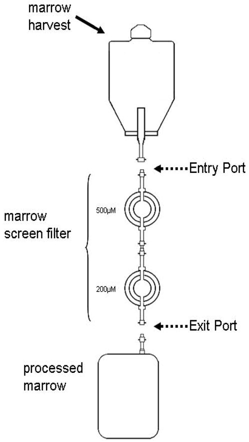
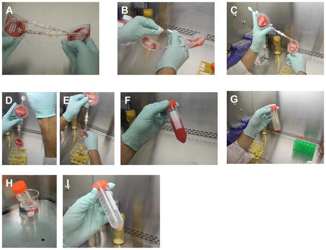
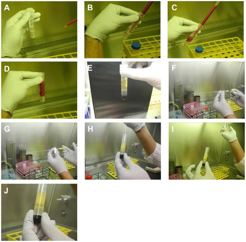
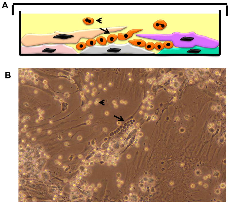
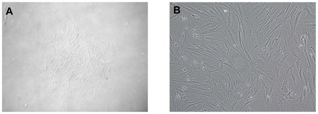
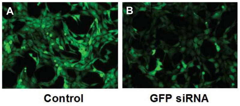
Marrow stromal cells (MSCs) are relatively rare cells difficult to visualize in marrow biopsies or detect in aspirated marrow. Under specific conditions MSC can be expanded in vitro and the population can give rise to several mesenchymal lineages. "MSC" also refers to mesenchymal stem cells which implies that all cells in the population are multipotent. It is generally agreed that while there may be a few multipotent stem cells in an MSC population the majority are not stem cells. In either case MSCs do not produce hematopoietic cells. Although MSCs have been isolated and characterized from several tissues, bone marrow is their most common source for research and clinical use. Primary MSC populations can be derived from bone marrow mononuclear cells with relative ease, but it is important to recognize the cellular heterogeneity within a culture and how this may vary from donor to donor. In this chapter, we describe methodology to derive primary MSCs from bone marrow screens, an otherwise discarded by-product of bone marrow harvests used for clinical transplantation. We also describe some useful techniques to characterize and manipulate MSCs-both primary and immortalized cell lines.


Figures

Figure 1
Figure 1. Schematic of Bone Marrow Harvest and Screens
The bone marrow aspirate collected from donors is hooked up typically to two filters called screens connected in series ( 500 μM first followed by 200 μM) to filter out particulate matter including spicules, clots and tissue fragments. The filtrate is collected for infusion ( processed marrow). The marrow screens are then made available to research laboratories with the entry port and exit connected to each other.
Figure 2
Figure 2. Extraction of bone marrow mononuclear cells (BMMNC) from marrow screens
A: Screens are typically provided with the two filters (500 μM and 200 μM) connected in series as a sterile loop. B. Carefully disengage the entrance port of the 500 μM filter from the exit port of the 200 μM filter. C: Pour any liquid contents left in the screen to a 50 cc conical tube. D: Inject 25 cc of PBS-EDTA into the entrance port of the 500 μM filter and collect the run through carefully. E: Try to dislodge any particulate material in the filters into the PBS-EDTA solution F: Approximately 50 cc of bone marrow particular material is eluted into a 50 cc conical tube. G: The cell pellet after the first wash is rich with erythroid cells which need to be mostly removed before plating in culture. H: Hemolysis of collected cells in hemolysis buffer at 37 degrees. I: Post-hemolysis and a further wash in PBS-EDTA, cell pellet is pale and has little visible erythroid contamination.
Figure 3
Figure 3. Extraction of bone marrow mononuclear cells (BMMNC) by ficoll gradient separation
A: 3ml of ficoll at room temperature is aliquoted in to 15 cc Falcon tubes B–D: 6–8 mf of dilute bone marrow is carefully layered on the ficoll layer taking care not to mix the two layers up. E: After centrifugation at 400 g for 30 minutes at room temperature, different layers become apparent. The mononuclear cells are trapped in the layer between ficoll and serum. F–J: With a sterile glass pipetted (stuffed with cotton at proximal end for seal), the mononuclear layer is carefully collected minimizing suction of both the ficoll layer between and the serum layer above.
Figure 4
Figure 4. Primary Long Term Cultures (LTCs or Dexter Cultures)
A: Cartoon depicting vertical layer of an LTC with stromal and hematopoietic elements. B: Phase contrast micrograph of an LTC. Long arrows in both panels depict the cobble stone areas (CSAs) comprised of primitive hematopoietic precursors trapped within the stromal layers and appear as “phase-dark” cells resembling a cobble-stone. Mature myeloid cells are released into the supernatant when the more primitive precursor cells in the CSAs divide and mature ( short arrow in both panels) and appear as “phase-light cells” in phase-contrast micrographs.
Figure 5
Figure 5. Morphology of MSC cultures
A: a Typical CFU-F that arises out of a single stromal precursor after 5–7 days of culture. B: Confluent cultures that develop after 2 weeks or more of culture.
Figure 6
Figure 6. CD146 expression in primary stromal cells and its use in sorting stromal cells
A and B: Immune Histochemistry (IHC) for CD146 expression of normal human bone marrow ( Panel A is an isotype antibody control. CD146 positive cells are present in a perivascular distribution, a location consistent with other models of where the HSPC niche might reside. C: Flow-cytometry analysis of MSC cultures after 1 passage ( 10–14 days of culture). A variable proportion of cells are CD146 positive and they inhabit of continuum of antigen expression. Two MSCs set up from separate donors are shown. D: Typical flow-sorting results from CD146-based sorting of MSCs. Approximately 35% of cells are deemed CD146 positive ( pre-sort, top histogram), and sorted to negative ( middle histogram) and CD146 negative ( bottom histogram) using a FACS-ARIA sorter ( Beckton Dickinson and Company) with a 100 μM nozzle.
Figure 7
Figure 7. Reverse Transfection of stromal cell lines
A: Stromal cell line HS27a stably expressing Green Fluorescent Protein (GFP) was transfected with control scrambled siRNA and visualized by inverted fluorescent microscopy after 48 hours. B: Hs27a-GFP cell lines transfected with anti-GFP siRNA showing marked reduction in GFP expression of most cells.

References

    1. Chabannon C, Torok-Storb B. Stem cell-stromal cell interactions. Curr Top Microbiol Immunol. 1992;177:123–36. - PubMed
    1. Trentin JJ. Determination of bone marrow stem cell differentiation by stromal hemopoietic inductive microenvironments (HIM) Am J Pathol. 1971;65:621–8. - PMC - PubMed
    1. McCulloch EA, Siminovitch L, Till JE, Russell ES, Bernstein SE. The cellular basis of the genetically determined hemopoietic defect in anemic mice of genotype sl-sld. Blood. 1965;26:399–410. - PubMed
    1. Huang E, Nocka K, Beier DR, Chu TY, Buck J, Lahm HW, et al. The hematopoietic growth factor KL is encoded by the sl locus and is the ligand of the c-kit receptor, the gene product of the W locus. Cell. 1990;63:225–33. - PubMed
    1. Williams DE, Eisenman J, Baird A, Rauch C, Van Ness K, March CJ, et al. Identification of a ligand for the c-kit proto-oncogene. Cell. 1990;63:167–74. - PubMed

Publication types