. 2013 Sep 27;288(39):27812-24.
doi: 10.1074/jbc.M113.465088. Epub 2013 Aug 19.

Dynein interacts with the neural cell adhesion molecule (NCAM180) to tether dynamic microtubules and maintain synaptic density in cortical neurons

Affiliations

Dynein interacts with the neural cell adhesion molecule (NCAM180) to tether dynamic microtubules and maintain synaptic density in cortical neurons

Eran Perlson et al. J Biol Chem. .

Abstract

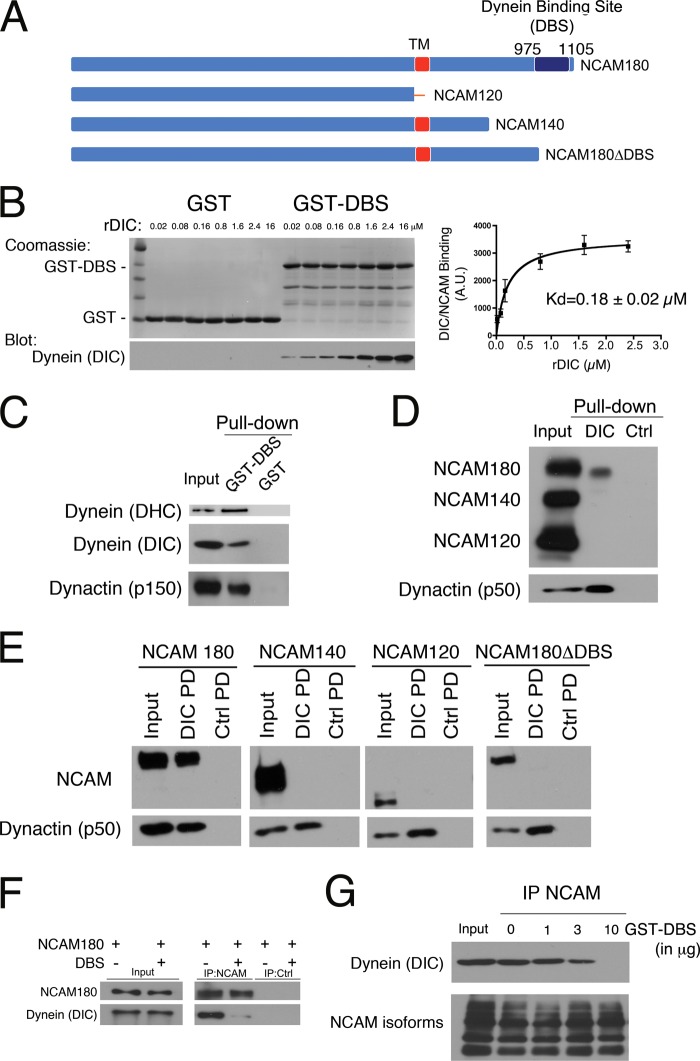
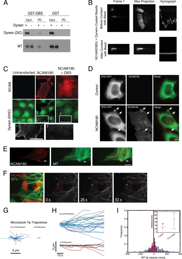
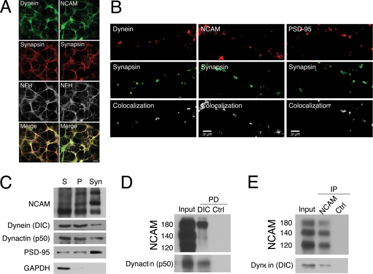
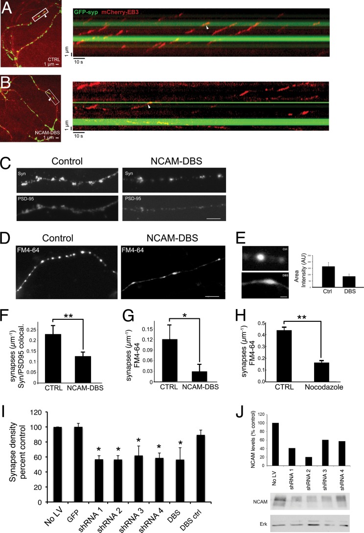
Cytoplasmic dynein is well characterized as an organelle motor, but dynein also acts to tether and stabilize dynamic microtubule plus-ends in vitro. Here we identify a novel and direct interaction between dynein and the 180-kDa isoform of the neural cell adhesion molecule (NCAM). Optical trapping experiments indicate that dynein bound to beads via the NCAM180 interaction domain can tether projecting microtubule plus-ends. Live cell assays indicate that the NCAM180-dependent recruitment of dynein to the cortex leads to the selective stabilization of microtubules projecting to NCAM180 patches at the cell periphery. The dynein-NCAM180 interaction also enhances cell-cell adhesion in heterologous cell assays. Dynein and NCAM180 co-precipitate from mouse brain extract and from synaptosomal fractions, consistent with an endogenous interaction in neurons. Thus, we examined microtubule dynamics and synaptic density in primary cortical neurons. We find that depletion of NCAM, inhibition of the dynein-NCAM180 interaction, or dampening of microtubule dynamics with low dose nocodazole all result in significantly decreased in synaptic density. Based on these observations, we propose a working model for the role of dynein at the synapse, in which the anchoring of the motor to the cortex via binding to an adhesion molecule mediates the tethering of dynamic microtubule plus-ends to potentiate synaptic stabilization.

Keywords: Cytoskeleton; Dynamic Instability; Dynein; Microtubules; NCAM; Synapses; Synaptosomes.


Figures

FIGURE 1.
FIGURE 1.
A direct interaction between dynein and NCAM180 recruits dynein to the cell cortex. A, three major isoforms of NCAM are expressed in mammalian neurons: NCAM180, NCAM140, and NCAM120. The DBS identified in the yeast two-hybrid screen, spanning residues 975–1105 of NCAM180, is shown. TM, transmembrane domain. B, direct binding of recombinant DIC to the DBS domain of NCAM180. Pull-down assays were performed with either GST or GST-DBS incubated with increasing concentrations of purified recombinant DIC (rDIC), as noted. GST-DBS and recombinant DIC bind with an apparent Kd of 0.18 ± 0.02 μm. C, dynein and dynactin are precipitated from mouse brain lysate by recombinant GST-DBS but not by GST. Input, 10% of total; Western blots were probed with antibodies to DHC, DIC, and dynactin (p150Glued subunit). D, dynein interacts specifically with NCAM180. NCAM180, but not NCAM140 or NCAM120, was precipitated from mouse brain extract with recombinant DIC bound to beads. Dynactin (p50 subunit) served as a positive control, and BSA control beads served as a negative control. Input, 10% of total. E, DIC pull-down (PD) experiments from COS7 cells transfected with mouse isoforms NCAM180, NCAM140, NCAM120, or with a construct lacking the dynein-binding site, NCAM180ΔDBS, demonstrate the specificity of the dynein-NCAM180 interaction and its dependence on the DBS domain. Input (10% of total), DIC pull-down (DIC PD), and BSA control pull-down (Ctrl PD) samples were probed with antibodies to NCAM; dynactin (p50) served as a positive control. F, co-expression of the DBS construct disrupts the co-immunoprecipitation of endogenous dynein with NCAM180 from COS7 cells; a control immunoprecipitation with protein A beads alone (IP:Ctrl) is also shown. G, disruption of the dynein-NCAM180 interaction is dose-dependent. The co-immunoprecipitation (IP) of endogenous dynein with NCAM from brain lysate is inhibited by the addition of purified recombinant GST-DBS (0, 1, 3, and 10 μg of DBS to 1 mg of total protein in lysate). Input, 10% of total. Error bars, S.E.
FIGURE 2.
FIGURE 2.
NCAM180 and dynein tether microtubule plus-ends in vitro and in cells. A, GST-DBS-bound beads or GST-bound control beads were incubated with purified microtubules in the presence (+) or absence (−) of dynein; binding was evaluated by Western blot with antibodies to dynein (DIC) and tubulin (MT). Input, 10% of total. Both dynein and microtubules bound to GST-DBS beads but not to control beads. B, an optical trap was used to position a DBS-dynein-bound bead near the diffusing plus-end of a microtubule, which was bound to the coverglass at its minus-end. A marked decrease in the lateral diffusion of the microtubule plus-end was observed after contact with the bead, as shown in both the maximum projection and the kymograph showing microtubule end dynamics over time. C, expression of exogenous NCAM180 in COS7 cells recruits dynein to the cell cortex (NCAM180). Co-expression of the DBS construct abrogates recruitment of dynein to the cortex. Insets are shown below at increased magnification. Scale bars, 25 μm. D, expression of NCAM180 in HeLa cells stably expressing DHC-GFP induces the recruitment of dynein to NCAM180 patches at the cortex (arrows); this cortical localization is not observed in untransfected control cells. Scale bar, 10 μm. E, microtubules project to the cell cortex and are transiently stabilized at NCAM patches localized to the cell surface in COS7 cells expressing NCAM180 (red, RFP-NCAM180; green, GFP-EMTB, a microtubule-associated protein used to report microtubule dynamics). F, live cell imaging of microtubule dynamics in COS7 cells expressing NCAM180 (see supplemental Videos 1–3). An initial two-color image (red, RFP-NCAM180; green, GFP-EMTB) shows the tethering of a microtubule plus-end at an NCAM180 patch (left). A time series shows the tethering over time of the microtubule plus-end (white dot) at a cortical NCAM180 patch (outlined in red) in COS7 cells. Note the robust dynamics of untethered microtubules (colored dots and lines) within the same cell; lines indicate microtubule tracks over time, and dots indicate the initial position. G, microtubule tip trajectories away from (left) and at NCAM patches (right) show the dampened dynamics characteristic of tethered microtubules. Trajectories are aligned such that the initial microtubule tip position is at the origin. H, dampening of microtubule tip dynamics at NCAM patches. Tracking the tip positions over time of microtubules away from (blue and green tracks, top) and in NCAM patches (red, yellow, and orange tracks, bottom) shows the altered dynamics of microtubule growth and shortening of tethered microtubules. Dots, time points where the microtubule tip was localized in an NCAM patch. I, histogram of the microtubule tip velocities at (red) and away from (blue) NCAM180 patches at the cortex. The inset shows the mean and 25 and 75% quartiles of the absolute values of the velocity of the microtubule plus-ends (p < 0.05; ANOVA).
FIGURE 3.
FIGURE 3.
Cell-cell interactions are dependent on microtubule dynamics and the dynein-NCAM interaction. A and B, COS7 cells were singly transfected with GFP, NCAM180, or NCAMΔDBS or doubly transfected with NCAM180 and the dynein inhibitor CC1. The enhanced aggregation induced by NCAM180 (p < 0.05) was not seen when the DBS was deleted or when dynein was inhibited by co-expression of the dominant negative construct CC1 (44). C, COS7 cells were transfected with NCAM180 and then treated either with either low dose (100 nm) or high dose (10 μm) nocodazole for 40 min. The enhanced aggregation induced by NCAM180 expression was blocked by nocodazole treatment to either depolymerize cellular microtubules at high dose nocodazole or dampen microtubule dynamics at low dose nocodazole. The percentage of cells in aggregates was scored relative to control cells expressing GFP. Error bars, S.E. from ≥3 replicates.
FIGURE 4.
FIGURE 4.
Dynein and NCAM both localize to synaptic sites and co-precipitate from synaptosomal fractions. A, dynein (green, left) and NCAM (green, right) localize with synapsin (red) in cortical neurons in contrast to neurofilament-H staining. Scale bar, 50 μm. B, dynein (red, left) and NCAM (red, center) both localize to synaptic sites along with the presynaptic marker synapsin shown in green, in the processes of cortical neurons at 14 DIV. The colocalization of postsynaptic marker PSD-95 (red, panels on right) with synapsin (green) is shown for comparison. Scale bar, 10 μm. The percentage of colocalization was determined from thresholded pre- and postsynaptic images using Imaris software (41); representative signals are shown (Colocalization) for each pair below the immunostained images. C, NCAM and dynein co-purify with synaptosomes. All three major NCAM isoforms (NCAM180, -140, and -120) were found in isolated synaptosomes, along with dynein (DIC) and dynactin (p50). Supernatant (S) and pellet (P) fractions from brain homogenate and the synaptosome-enriched fraction (Syn) were probed for the postsynaptic marker PSD-95, which is selectively enriched in the synaptosome fraction, and the cytosolic marker GAPDH, which is depleted. D and E, pull-down experiments from mouse brain synaptosomes show that NCAM180 is precipitated by recombinant DIC-bound beads (D) and that endogenous dynein is co-immunoprecipitated with NCAM antibodies (E).
FIGURE 5.
FIGURE 5.
Inhibition of the dynein-NCAM180 interaction disrupts microtubule dynamics and decreases synaptic density in primary cortical neurons. A, microtubule dynamics at synapses. Cortical neurons were transfected with EB3-mCherry and GFP-synaptophysin to image microtubule dynamics and synaptic sites. Shown in the left panel is a representative image from a single time point, and shown on the right is a kymograph generated from a line drawn along a neuronal process in the time series. A white arrowhead denotes the same representative EB3 comet in both the micrograph and the kymograph. These data show that EB3 comets preferentially terminate at synaptic sites in control neurons. B, expression of DBS alters microtubule dynamics at synapses. In neurons expressing the DBS from NCAM180, EB3 comets no longer terminate preferentially at synaptic sites. A representative micrograph is shown on the left, and a kymograph from the time series is shown on the right, with the same comet tail indicated in both panels by a white arrowhead. Expression of the NCAM-DBS domain reduces the fraction of EB3 comets terminating at synapses (n > 85 comets in >7 neurons). C, expression of the DBS from NCAM180 reduces synaptic density in cortical neurons, as assessed by colocalization of puncta positive for both pre- and postsynaptic markers (synapsin (top) and PSD-95 (bottom)). D, expression of the DBS from NCAM180 reduces synaptic density in cortical neurons as assessed by measuring KCl-stimulated uptake of FM4-64. E, expression of the NCAM-DBS in cortical neurons disrupts the appearance of FM4-64-positive synaptic sites. Synapses from cortical neurons transfected with the DBS from NCAM180 have a decreased area intensity (DBS; 86 ± 19 arbitrary units (A.U.)) compared with control neurons (Ctrl; 164 ± 31 arbitrary units, p < 0.05). Synapses in DBS-transfected neurons are also more broad, 2.4 ± 0.6 μm compared with 1.8 ± 0.4 μm in control neurons (FWHM; p < 0.05). F and G, quantitative decreases in synaptic density induced by expression of DBS assessed using either colocalization of pre- and postsynaptic markers (p < 0.01, n = 15 neurons with >1000 synapses from three experiments; error bars, S.E.) (F) or FM4-64 uptake (p < 0.01, n = 18 neurons with >1000 synapses from six experiments) (G). H, dampening microtubule dynamics decreased synaptic density. The effects of DBS expression on synaptic density can be compared with the effects of treatment of cortical neurons with low dose nocodazole (50 nm for 20 h) (p < 0.01, n = 27 neurons with >500 synapses from two experiments). Experimental values were measured relative to same day controls because synaptic density is dependent on plating density and DIV. I, depletion of NCAM using four different shRNA (shRNA 1–4) constructs or expression of the dominant negative DBS construct resulted in similar decreases in synaptic density, relative to controls (NO LV, no lentivirus infection; GFP, infection with lentivirus expressing GFP; DBS ctrl, mock transfection). For all panels, *, p < 0.05; **, p < 0.01. J, depletion of NCAM isoforms from cortical neuronal cultures by lentivirus expression of shRNA constructs targeting NCAM as assessed by Western blot; ERK immunoreactivity was used as a loading control.

Similar articles

Cited by

References

    1. Kardon J. R., Vale R. D. (2009) Regulators of the cytoplasmic dynein motor. Nat. Rev. Mol. Cell Biol. 10, 854–865 - PMC - PubMed
    1. Nyarko A., Song Y., Barbar E. (2012) Intrinsic disorder in dynein intermediate chain modulates its interactions with NudE and dynactin. J. Biol. Chem. 287, 24884–24893 - PMC - PubMed
    1. Kirschner M., Mitchison T. (1986) Beyond self-assembly. From microtubules to morphogenesis. Cell 45, 329–342 - PubMed
    1. Levy J. R., Holzbaur E. L. (2007) Special delivery. Dynamic targeting via cortical capture of microtubules. Dev. Cell 12, 320–322 - PubMed
    1. Heil-Chapdelaine R. A., Oberle J. R., Cooper J. A. (2000) The cortical protein Num1p is essential for dynein-dependent interactions of microtubules with the cortex. J. Cell Biol. 151, 1337–1344 - PMC - PubMed

Publication types