

Decreasing tropomyosin phosphorylation rescues tropomyosin-induced familial hypertrophic cardiomyopathy
- PMID: 23960072
- PMCID: PMC3789987
- DOI: 10.1074/jbc.M113.466466
Decreasing tropomyosin phosphorylation rescues tropomyosin-induced familial hypertrophic cardiomyopathy
Abstract
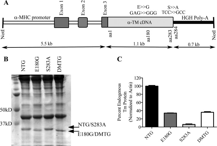
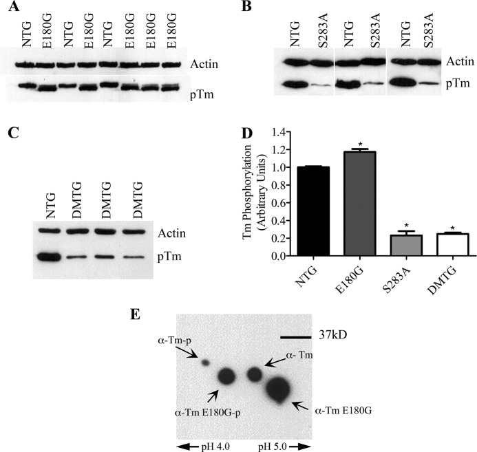
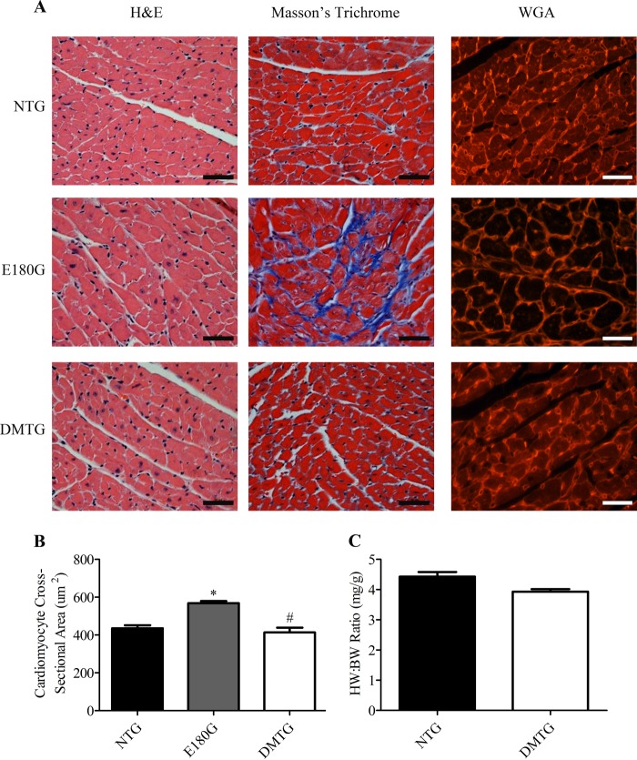
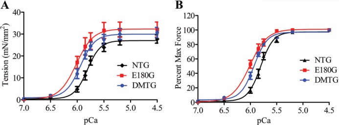
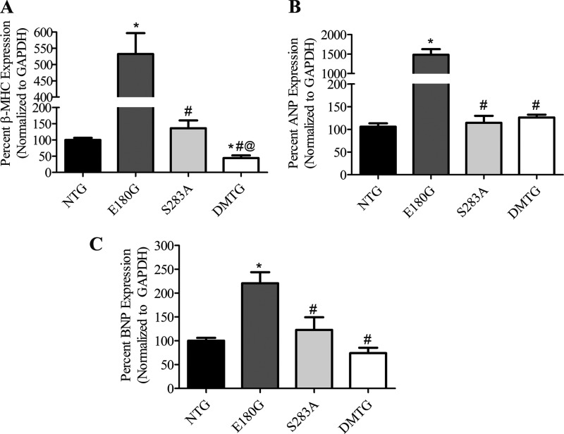
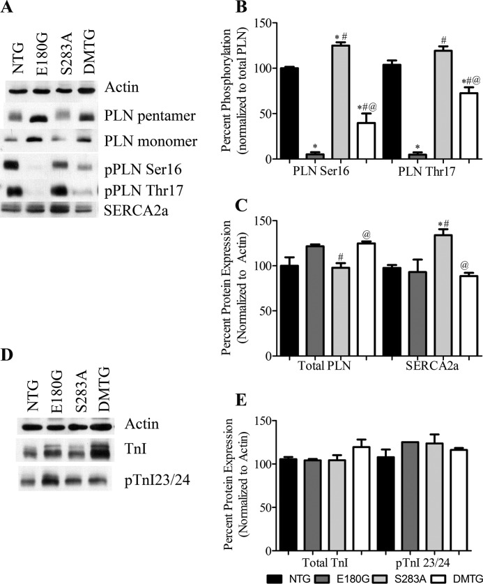
Studies indicate that tropomyosin (Tm) phosphorylation status varies in different mouse models of cardiac disease. Investigation of basal and acute cardiac function utilizing a mouse model expressing an α-Tm protein that cannot be phosphorylated (S283A) shows a compensated hypertrophic phenotype with significant increases in SERCA2a expression and phosphorylation of phospholamban Ser-16 (Schulz, E. M., Correll, R. N., Sheikh, H. N., Lofrano-Alves, M. S., Engel, P. L., Newman, G., Schultz Jel, J., Molkentin, J. D., Wolska, B. M., Solaro, R. J., and Wieczorek, D. F. (2012) J. Biol. Chem. 287, 44478-44489). With these results, we hypothesized that decreasing α-Tm phosphorylation may be beneficial in the context of a chronic, intrinsic stressor. To test this hypothesis, we utilized the familial hypertrophic cardiomyopathy (FHC) α-Tm E180G model (Prabhakar, R., Boivin, G. P., Grupp, I. L., Hoit, B., Arteaga, G., Solaro, R. J., and Wieczorek, D. F. (2001) J. Mol. Cell. Cardiol. 33, 1815-1828). These FHC hearts are characterized by increased heart:body weight ratios, fibrosis, increased myofilament Ca(2+) sensitivity, and contractile defects. The FHC mice die by 6-8 months of age. We generated mice expressing both the E180G and S283A mutations and found that the hypertrophic phenotype was rescued in the α-Tm E180G/S283A double mutant transgenic animals; these mice exhibited no signs of cardiac hypertrophy and displayed improved cardiac function. These double mutant transgenic hearts showed increased phosphorylation of phospholamban Ser-16 and Thr-17 compared with the α-Tm E180G mice. This is the first study to demonstrate that decreasing phosphorylation of tropomyosin can rescue a hypertrophic cardiomyopathic phenotype.
Keywords: Calcium; Cardiac Hypertrophy; Muscle; Phosphorylation; Transgenic Mice; Tropomyosin.
Figures
Similar articles
-
Alpha-tropomyosin mutations in inherited cardiomyopathies.J Muscle Res Cell Motil. 2013 Aug;34(3-4):285-94. doi: 10.1007/s10974-013-9358-5. Epub 2013 Sep 5. J Muscle Res Cell Motil. 2013. PMID: 24005378 Review.
-
Cardiac dysfunction in hypertrophic cardiomyopathy mutant tropomyosin mice is transgene-dependent, hypertrophy-independent, and improved by beta-blockade.Circ Res. 2002 Aug 9;91(3):255-62. doi: 10.1161/01.res.0000027530.58419.82. Circ Res. 2002. PMID: 12169652
-
Rescue of tropomyosin-induced familial hypertrophic cardiomyopathy mice by transgenesis.Am J Physiol Heart Circ Physiol. 2007 Aug;293(2):H949-58. doi: 10.1152/ajpheart.01341.2006. Epub 2007 Apr 6. Am J Physiol Heart Circ Physiol. 2007. PMID: 17416600
-
N-acetylcysteine reverses diastolic dysfunction and hypertrophy in familial hypertrophic cardiomyopathy.Am J Physiol Heart Circ Physiol. 2015 Nov 15;309(10):H1720-30. doi: 10.1152/ajpheart.00339.2015. Epub 2015 Oct 2. Am J Physiol Heart Circ Physiol. 2015. PMID: 26432840 Free PMC article.
-
Cardiac myosin binding protein C phosphorylation in cardiac disease.J Muscle Res Cell Motil. 2012 May;33(1):43-52. doi: 10.1007/s10974-011-9280-7. Epub 2011 Nov 30. J Muscle Res Cell Motil. 2012. PMID: 22127559 Free PMC article. Review.
Cited by
-
Genetically Encoded Biosensors Reveal PKA Hyperphosphorylation on the Myofilaments in Rabbit Heart Failure.Circ Res. 2016 Sep 30;119(8):931-43. doi: 10.1161/CIRCRESAHA.116.308964. Epub 2016 Aug 30. Circ Res. 2016. PMID: 27576469 Free PMC article.
-
Analysis of fibrosis in control or pressure overloaded rat hearts after mechanical unloading by heterotopic heart transplantation.Sci Rep. 2019 Apr 5;9(1):5710. doi: 10.1038/s41598-019-42263-1. Sci Rep. 2019. PMID: 30952943 Free PMC article.
-
Alpha-tropomyosin mutations in inherited cardiomyopathies.J Muscle Res Cell Motil. 2013 Aug;34(3-4):285-94. doi: 10.1007/s10974-013-9358-5. Epub 2013 Sep 5. J Muscle Res Cell Motil. 2013. PMID: 24005378 Review.
-
Phenotyping cardiomyopathy in adult zebrafish.Prog Biophys Mol Biol. 2018 Oct;138:116-125. doi: 10.1016/j.pbiomolbio.2018.05.013. Epub 2018 May 30. Prog Biophys Mol Biol. 2018. PMID: 29884423 Free PMC article. Review.
-
Hypertrophic Cardiomyopathy: A Vicious Cycle Triggered by Sarcomere Mutations and Secondary Disease Hits.Antioxid Redox Signal. 2019 Aug 1;31(4):318-358. doi: 10.1089/ars.2017.7236. Epub 2018 Apr 11. Antioxid Redox Signal. 2019. PMID: 29490477 Free PMC article. Review.
References
Publication types
MeSH terms
Substances
Grants and funding
LinkOut - more resources
Full Text Sources
Other Literature Sources
Molecular Biology Databases
Miscellaneous
