. 2013 Oct 4;288(40):28535-47.
doi: 10.1074/jbc.M113.508085. Epub 2013 Aug 19.

JNK3 enzyme binding to arrestin-3 differentially affects the recruitment of upstream mitogen-activated protein (MAP) kinase kinases

Affiliations

JNK3 enzyme binding to arrestin-3 differentially affects the recruitment of upstream mitogen-activated protein (MAP) kinase kinases

Xuanzhi Zhan et al. J Biol Chem. .

Abstract

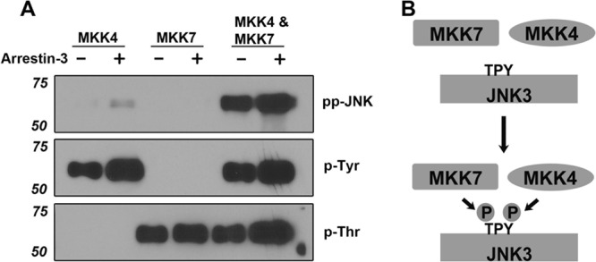
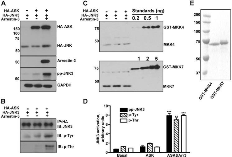
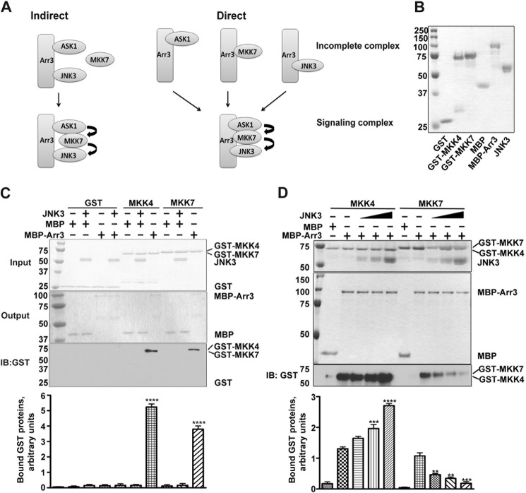
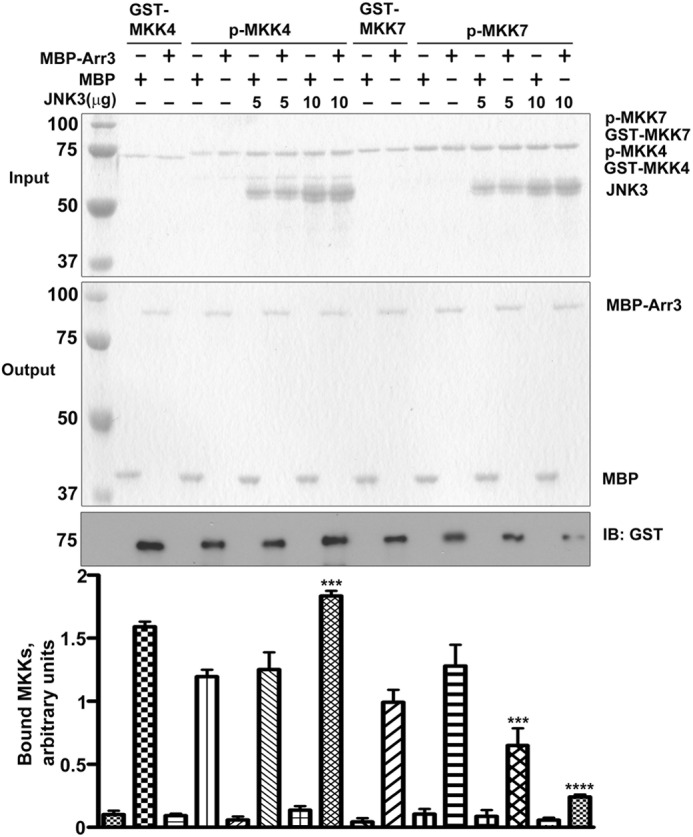
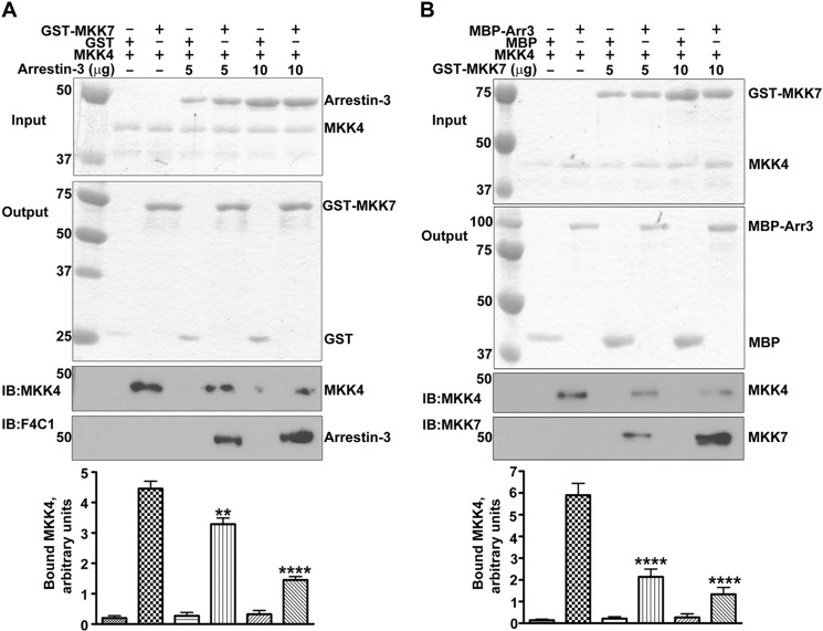
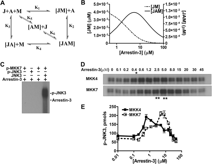
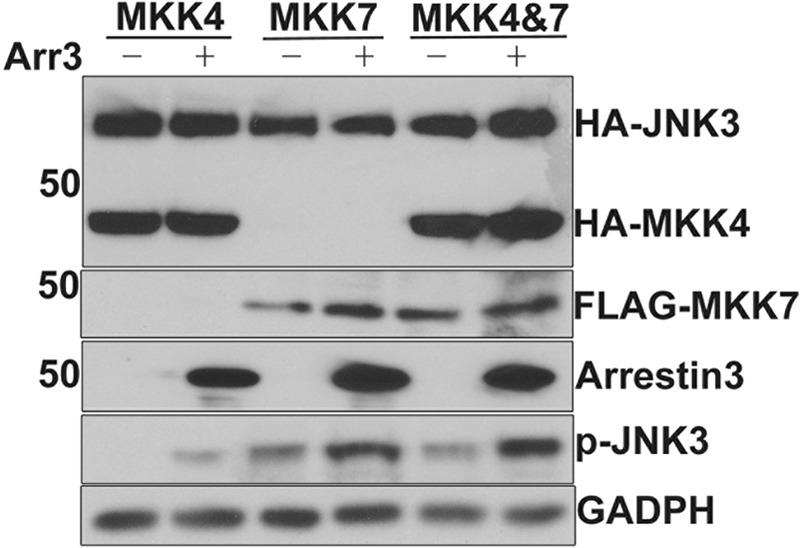
Arrestin-3 was previously shown to bind JNK3α2, MKK4, and ASK1. However, full JNK3α2 activation requires phosphorylation by both MKK4 and MKK7. Using purified proteins we show that arrestin-3 directly interacts with MKK7 and promotes JNK3α2 phosphorylation by both MKK4 and MKK7 in vitro as well as in intact cells. The binding of JNK3α2 promotes an arrestin-3 interaction with MKK4 while reducing its binding to MKK7. Interestingly, the arrestin-3 concentration optimal for scaffolding the MKK7-JNK3α2 module is ∼10-fold higher than for the MKK4-JNK3α2 module. The data provide a mechanistic basis for arrestin-3-dependent activation of JNK3α2. The opposite effects of JNK3α2 on arrestin-3 interactions with MKK4 and MKK7 is the first demonstration that the kinase components in mammalian MAPK cascades regulate each other's interactions with a scaffold protein. The results show how signaling outcomes can be affected by the relative expression of scaffolding proteins and components of signaling cascades that they assemble.

Keywords: Arrestin; Cell Signaling; Jun N-terminal Kinase (JNK); MAP Kinases (MAPKs); Protein Kinases; Protein Phosphorylation.


Figures

FIGURE 1.
FIGURE 1.
The specificity of MKK4 and MKK7 toward JNK3α2 is not affected by arrestin-3. A, Western blot of MKK-phosphorylated JNK3α2. In the presence (+) or absence (−) of arrestin-3 (1 μm), 1 μm JNK3α2 was incubated with active MKK4, MKK7, or MKK4+MKK7 combination for 3 min. The reactions were stopped by SDS sample buffer and analyzed by Western blot with phospho-JNK3α2 (ppJNK), phosphotyrosine (p-Tyr), and phosphothreonine (p-Thr) antibodies, as indicated. B, a schematic showing that full activation of JNK3α2 requires both MKK4 and MKK7 that phosphorylate Tyr-223 and Thr-221, respectively.
FIGURE 2.
FIGURE 2.
The phosphorylation of both tyrosine and threonine on JNK3α2 is enhanced by arrestin-3. A, COS-7 cells were transfected with JNK3α2 alone or in combination with HA-ASK1 with or without arrestin-3. Cell lysates were analyzed by Western blot using indicated antibodies. GADPH served as the loading control. B, HA-JNK3α2 in cell lysates was immunoprecipitated (IP) by an anti-HA antibody. The immunoprecipitate was analyzed by Western blot (IB) using anti-HA, Tyr(P) (p-Tyr), or Thr(P) (p-Thr) antibodies. C, cell lysates containing 10 or 5 μg of total protein were used to quantify endogenous MKK4 and MKK7, respectively, by Western blot with antibodies recognizing MKK4 (upper panel) or MKK7 (lower panel). Purified GST-MKK4 (0.2, 0.5, 1 ng) and GST-MKK7 (1, 2, 5 ng) were used as standards. D, quantitative analysis of arrestin-3 effect on JNK3α2 phosphorylation (representative blots shown in A (ppJNK) and B (Tyr(P) and Thr(P)). Means ± S.D. (n = 3) of relative intensity of phospho-JNK3 (***, p < 0.001, as compared with the basal level in cells co-expressing ASK1 and JNK3 without arrestin-3), Tyr(P) (**, p < 0.01), and Thr(P) (***, p < 0.001) bands are shown. E, Coomassie Blue-stained SDS-PAGE gel showing the purity of GST-MKK4 and GST-MKK7 used as standards to generate calibration curves (1 μg of each protein was loaded).
FIGURE 3.
FIGURE 3.
JNK3α2 has an opposite effect on arrestin-3 binding to MKK4 and MKK7. A, two proposed models of arrestin-3-mediated scaffolding of ASK1-MKK7-JNK3 cascade. Indirect model (left panel), MKK7 is recruited to the complex through ASK1 and JNK3 without direct binding to arrestin-3. Direct model (right panel), arrestin-3 directly binds all three kinases to form a complete signaling complex. B, Coomassie-stained SDS-PAGE gel showing the purity of proteins used in the assays shown in C and D. C, an MBP pulldown assay showed that both MKK4 and MKK7 were retained by MBP-arrestin-3 but not by MBP (control). No nonspecific binding of GST to MBP-arrestin-3 or MBP was detected. Top panel, Coomassie Blue-stained SDS-PAGE gel showing the input of GST fusion proteins and JNK3α2. Middle panel, Coomassie Blue staining of MBP and MBP-Arr3 eluted from the amylose column. Bottom panels, Western blot (IB) of the eluates from the indicated columns and bar graph showing the quantification of the Western blot; means ± S.D. (n = 3) of the relative intensity of bands are shown (****, p < 0.0001, as compared with MBP control). Note that both GST-tagged MKK4 and MKK7 are retained by MBP-Arr3, but not by MBP, and no nonspecific effects of JNK3α2 on the pulldown were observed. D, JNK3α2 enhanced the binding of MKK4 but decreased the binding of MKK7 to arrestin-3. Top panel, Coomassie staining of input of the indicated proteins (10%). Middle panel, Coomassie staining of eluted proteins (20%). Bottom panel, Western blots of ⅕ of the eluate to detect the indicated GST-tagged proteins. The amounts of JNK3α2 in the assay were (left to right): 2, 5, and 10 μg. Representative gels are shown. Bar graph, quantification of the Western blot data. Means ± S.D. (n = 3) of the relative intensity of bands are shown (**, p < 0.01: ***, p < 0.001; ****, p < 0.0001, as compared with corresponding control binding in the absence of JNK3α2).
FIGURE 4.
FIGURE 4.
Arrestin-3 binding to active MKK4 and active MKK7 is regulated by JNK3α2. The binding of active MKKs to arrestin-3 was measured and compared with their inactive forms by MBP pulldown assay. The effects of JNK3α2 on the interaction of arrestin-3 with active MKK4 and 7 were also analyzed. Top panel, Coomassie Blue-stained gels of input of prey proteins (JNK3, GST-MKK4, active GST-MKK4 (p-MKK4), GST-MKK7, and active GST-MKK7 (p-MKK7). Middle panel, Coomassie Blue-stained gel of the eluted bait proteins (MBP and MBP-arrestin-3). Bottom panel, the amount of GST-tagged MKK4 or MKK7 retained was analyzed by Western blot. Representative gels are shown. Bar graph, quantification of the Western blot data. Means ± S.D. (n = 3) of the relative intensity of bands are shown (***, p < 0.001; ****, p < 0.0001, as compared with corresponding control binding in the absence of JNK3α2). IB, immunoblot.
FIGURE 5.
FIGURE 5.
MKK4 and MKK7 compete for arrestin-3 binding. A, pulldown of MKK4 by GST-MKK7 but not by GST (control). MKK4/MKK7 interaction was decreased by arrestin-3. Top panel, Coomassie Blue stained gel of input of prey proteins (arrestin-3 and MKK4). Middle panel, Coomassie Blue-stained gel of the output of bait proteins (GST and GST-MKK7). Lower panels, the amounts of retained MKK4 and arrestin-3 were analyzed by Western blot with indicated antibodies. Representative gels are shown. Bar graph, quantification of the MKK4 retained by GST-MKK7. Means ± S.D. (n = 3) of the relative intensity of bands (**, p < 0.01; ****, p < 0.0001, as compared with control without arrestin-3). IB, immunoblot. B, MKK4 and MKK7 compete for arrestin-3 binding. The amount of MKK4 retained by immobilized MBP-arrestin-3 progressively decreased in the presence of increasing concentrations of GST-MKK7. Coomassie Blue-stained gels of input of prey proteins (MKK4 and GST-MKK7) and output of bait proteins (MBP and MBP-Arr3) are shown in top two panels, respectively; the amount of MKK4 or GST-MKK7 retained was analyzed by Western blot using indicated antibodies. Representative gels are shown. Bar graph, quantification of the MKK4 retained by MBP-arrestin-3. Means ± S.D. (n = 3) of the relative intensity of bands (****, p < 0.0001, as compared with control without GST-MKK7).
FIGURE 6.
FIGURE 6.
Biphasic effects of arrestin-3 on JNK3α2 activation by MKK7 and MKK4. A, a model showing the scaffolding mechanism of the two-kinase signaling module. A, J, and M stand for arrestin-3, JNK3α2, and upstream kinase MKK4 or MKK7, respectively. Kinases can exist in three different complexes: (a) directly interacting JM; (b) bound to the scaffold to form incomplete complexes containing a single kinase (JA or AM); (c) simultaneously tethered by a scaffold to form a complete two-kinase signaling complex JAM. Six affinity constants (K1--K6) describe the indicated binding equilibria. B, calculated concentrations of JM (dotted line, left y axis) and JAM (solid line, right y axis) complexes (KinTek Explorer 3.0; all six Kd values were set at 5 μm). C, active MKK7 and JNK3α2 do not phosphorylate arrestin-3. Purified arrestin-3 (1 μm) was incubated with active MKK7 (50 nm), active JNK3α2 (50 nm), or inactive JNK3α2 (0.5 μm) for 5 min. The reactions were stopped with SDS sample buffer, and the proteins were separated on 10% SDS-PAGE. The gel was dried and exposed to x-ray film. A representative autoradiogram from one of four experiments is shown. D, representative autoradiograms showing JNK3α2 phosphorylated by MKK4 (upper panel) and MKK7 (lower panel) at the indicated concentration of arrestin-3 (10-s incubation). The optimal arrestin-3 concentrations are indicated (*, for MKK4; **, for MKK7). E, the effect of arrestin-3 concentration on JNK3α2 phosphorylation by both MKK4 and MKK7 is biphasic. The bands from the autoratiogram gels were cut out, and the radioactivity was measured in a Tri-Carb liquid scintillation counter to quantify the incorporation of [32P]phosphate from [γ-32P]ATP into JNK3α2. Means ± S.D. (n = 3) are shown.
FIGURE 7.
FIGURE 7.
Arrestin-3 enhances the phosphorylation of JNK3α2 by MKK4 or MKK7 in intact cells. COS-7 cells were transfected with HA-JNK3α2 in combination with HA-MKK4, FLAG-MKK7, or both with (+) or without (−) arrestin-3. Cell lysates were analyzed by Western blot using the indicated antibodies. Note that co-expression of arrestin-3 enhances JNK3α2 phosphorylation by either MKK. GADPH served as the loading control.

References

    1. Gurevich V. V., Gurevich E. V. (2006) The structural basis of arrestin-mediated regulation of G-protein-coupled receptors. Pharmacol. Ther. 110, 465–502 - PMC - PubMed
    1. DeWire S. M., Ahn S., Lefkowitz R. J., Shenoy S. K. (2007) β-Arrestins and cell signaling. Annu. Rev. Physiol. 69, 483–510 - PubMed
    1. Gurevich E. V., Gurevich V. V. (2006) Arrestins. Ubiquitous regulators of cellular signaling pathways. Genome Biol. 7, 236. - PMC - PubMed
    1. Xiao K., McClatchy D. B., Shukla A. K., Zhao Y., Chen M., Shenoy S. K., Yates J. R., 3rd, Lefkowitz R. J. (2007) Functional specialization of β-arrestin interactions revealed by proteomic analysis. Proc. Natl. Acad. Sci. U.S.A. 104, 12011–12016 - PMC - PubMed
    1. Pearson G., Robinson F., Beers Gibson T., Xu B. E., Karandikar M., Berman K., Cobb M. H. (2001) Mitogen-activated protein (MAP) kinase pathways. Regulation and physiological functions. Endocr. Rev. 22, 153–183 - PubMed

Publication types