. 2013 Nov;84(5):687-95.
doi: 10.1124/mol.113.088096. Epub 2013 Aug 19.

Caged naloxone reveals opioid signaling deactivation kinetics

Affiliations

Caged naloxone reveals opioid signaling deactivation kinetics

Matthew R Banghart et al. Mol Pharmacol. 2013 Nov.

Abstract

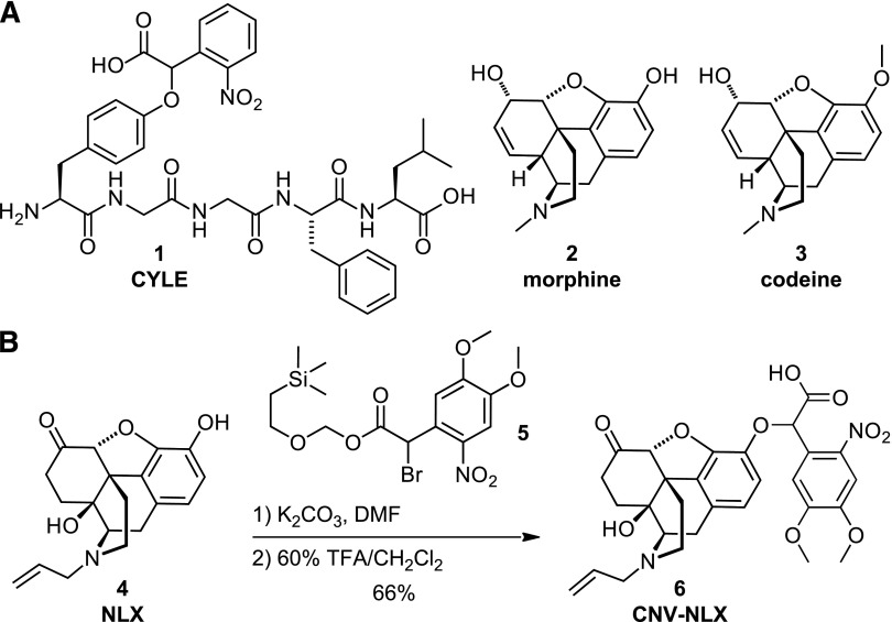
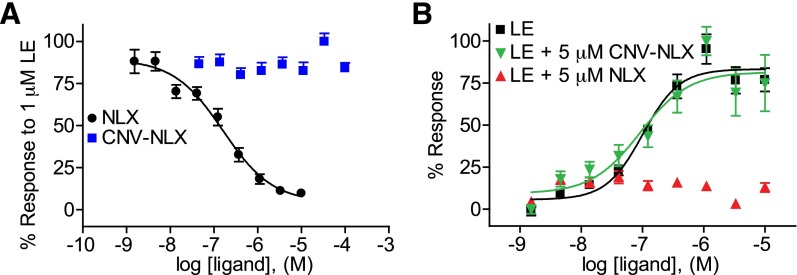
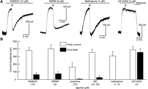
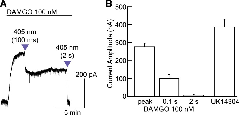
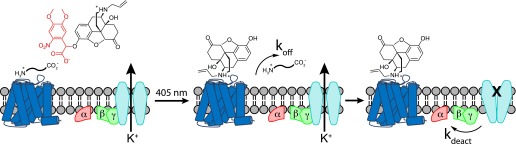
The spatiotemporal dynamics of opioid signaling in the brain remain poorly defined. Photoactivatable opioid ligands provide a means to quantitatively measure these dynamics and their underlying mechanisms in brain tissue. Although activation kinetics can be assessed using caged agonists, deactivation kinetics are obscured by slow clearance of agonist in tissue. To reveal deactivation kinetics of opioid signaling we developed a caged competitive antagonist that can be quickly photoreleased in sufficient concentrations to render agonist dissociation effectively irreversible. Carboxynitroveratryl-naloxone (CNV-NLX), a caged analog of the competitive opioid antagonist NLX, was readily synthesized from commercially available NLX in good yield and found to be devoid of antagonist activity at heterologously expressed opioid receptors. Photolysis in slices of rat locus coeruleus produced a rapid inhibition of the ionic currents evoked by multiple agonists of the μ-opioid receptor (MOR), but not of α-adrenergic receptors, which activate the same pool of ion channels. Using the high-affinity peptide agonist dermorphin, we established conditions under which light-driven deactivation rates are independent of agonist concentration and thus intrinsic to the agonist-receptor complex. Under these conditions, some MOR agonists yielded deactivation rates that are limited by G protein signaling, whereas others appeared limited by agonist dissociation. Therefore, the choice of agonist determines which feature of receptor signaling is unmasked by CNV-NLX photolysis.


Figures

Fig. 1.
Fig. 1.
Design and synthesis of CNV-NLX, a caged opioid antagonist. (A) Chemical structures of carboxynitrobenzyl-tyrosine-[Leu5]-enkephalin (CYLE) (1), morphine (2), and codeine (3). (B) Two-step synthesis of CNV-NLX (6) from NLX (4).
Fig. 2.
Fig. 2.
CNV-NLX is inactive at the μ-opioid receptor expressed in human embryonic kidney 293 cells. (A) Dose-response curves of NLX (black circles) and CNV-NLX (blue squares) in the presence of 1 μM LE (n = 12 wells). (B) Dose-response curves of LE alone (black squares), and in the presence of 5 μM NLX (upward red triangles) and CNV-NLX (downward green triangles) (n = 6 wells).
Fig. 3.
Fig. 3.
Light-driven antagonism of opioid receptors by CNV-NLX. (A) Photolysis inhibits currents evoked by high concentrations of the MOR agonists DAMGO, DERM, and methadone, but causes only an artifactual response at α-adrenergic receptors in the presence of UK14304. (B) Summary plot comparing currents evoked by MOR agonists and the α-adrenergic agonist UK14304, before and after CNV-NLX photolysis (n = 4–8 cells per agonist). Two concentrations were pooled for both ME and methadone. Purple triangles indicate 5-second flashes of 405-nm light.
Fig. 4.
Fig. 4.
Analog photocontrol of NLX release. (A) Varying the duration of the light flash produces graded inhibition of the outward current evoked by bath perfusion of a subsaturating concentration of DAMGO. (B) Summary plot comparing the current amplitudes produced by DAMGO (100 nM) before and after each uncaging stimulus to that produced by a saturating concentration of UK14304 (3 µM, n = 4 cells). Because DAMGO (100 nM) produced smaller peak currents than UK14304 (3 µM), the opioid stimulus is confirmed to be subsaturating.
Fig. 5.
Fig. 5.
Schematic illustrating the molecular events that underlie opioid signaling deactivation in response to antagonist uncaging in the presence of agonist. The photo-released competitive antagonist can bind only after agonist dissociation, which occurs with a rate constant of koff. In the agonist-free state, the G protein subunits that support GIRK channel opening dissociate from the activated channels, allowing channel closure with a rate constant of kdeact.
Fig. 6.
Fig. 6.
Low agonist concentrations reveal DERM ligand dissociation kinetics. (A) Superimposition of the currents evoked by 10 nM and 10 μM DERM (left); amplitude-normalized currents reveal slower deactivation at saturating agonist concentration (right). (B) Summary plot of the DERM current, normalized to the current induced by UK14304 (3 µM, black squares), and tau decay (open circles) as a function of DERM concentration (n = 4–8 cells per condition). The dashed line indicates the average of the time constants measured at 10, 30, and 100 nM, which did not vary significantly.
Fig. 7.
Fig. 7.
Light-induced deactivation of currents produced by various MOR agonists at subsaturating concentrations. (A) Superimposition of the normalized currents evoked by subsaturating concentrations of various small-molecule (left) and peptide (right) agonists. (B) Summary plot of the normalized currents (white bars) and corresponding light-induced deactivation rates (black bars) at the indicated subsaturating concentrations. Opioid agonist–induced peak currents are normalized to the peak currents produced by UK14303 (3 µM) in the same cells (n = 4–8 cells per condition). (C) Summary plot of the deactivation time constant versus agonist affinity according to compound class (n = 4–8 cells per condition).

References

    1. Albizu L, Balestre MN, Breton C, Pin JP, Manning M, Mouillac B, Barberis C, Durroux T. (2006) Probing the existence of G protein-coupled receptor dimers by positive and negative ligand-dependent cooperative binding. Mol Pharmacol 70:1783–1791 - PubMed
    1. Banghart MR, Sabatini BL. (2012) Photoactivatable neuropeptides for spatiotemporally precise delivery of opioids in neural tissue. Neuron 73:249–259 - PMC - PubMed
    1. Birdsong WT, Arttamangkul S, Clark MJ, Cheng K, Rice KC, Traynor JR, Williams JT. (2013) Increased agonist affinity at the μ-opioid receptor induced by prolonged agonist exposure. J Neurosci 33:4118–4127 - PMC - PubMed
    1. Childers SR, Snyder SH. (1980) Differential regulation by guanine nucleotides or opiate agonist and antagonist receptor interactions. J Neurochem 34:583–593 - PubMed
    1. Connor M, Traynor J. (2010) Constitutively active μ-opioid receptors. Methods Enzymol 484:445–469 - PubMed

Publication types

LinkOut - more resources