

Case Reports
doi: 10.1136/bcr-2013-200083.
Delayed postirradiation camptocormia
Affiliations
- PMID: 23960150
- PMCID: PMC3762404
- DOI: 10.1136/bcr-2013-200083
Item in Clipboard
Case Reports
Delayed postirradiation camptocormia
BMJ Case Rep.
.
Abstract
Radiotherapy may cause central or peripheral nervous system complications, reported examples being myelopathy, brachial/lumbosacral plexopathies or predominantly motor lumbosacral radiculopathy. In the literature some studies regard camptocormia as a paravertebral myopathy and others as of neurogenic origin. We present a patient who developed camptocormia, 42 years after radiation therapy to the para-aortic and inguinal area.
Figures
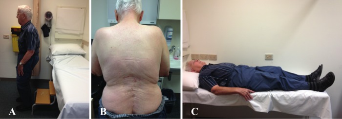
(A) Forward flexion of the spine compensated by bending of the knees when standing. (B) Marked atrophy of the paraspinal muscles. (C) Normal position of the spine when lying down.
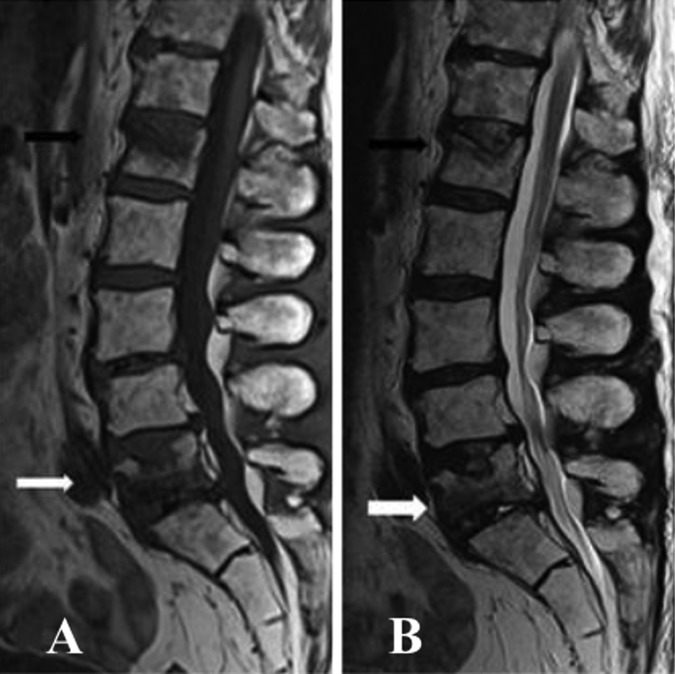
(A and B) Sagittal T2 and T1 images of the lumbar spine demonstrated increased signals throughout the lumbar vertebrae in keeping with postradiotherapy change. Mild to moderate reduction in height of L1 vertebral body (white arrow) and L5 vertebral body (black arrow) with superior end-plate involvement would be the result of old compression fractures.
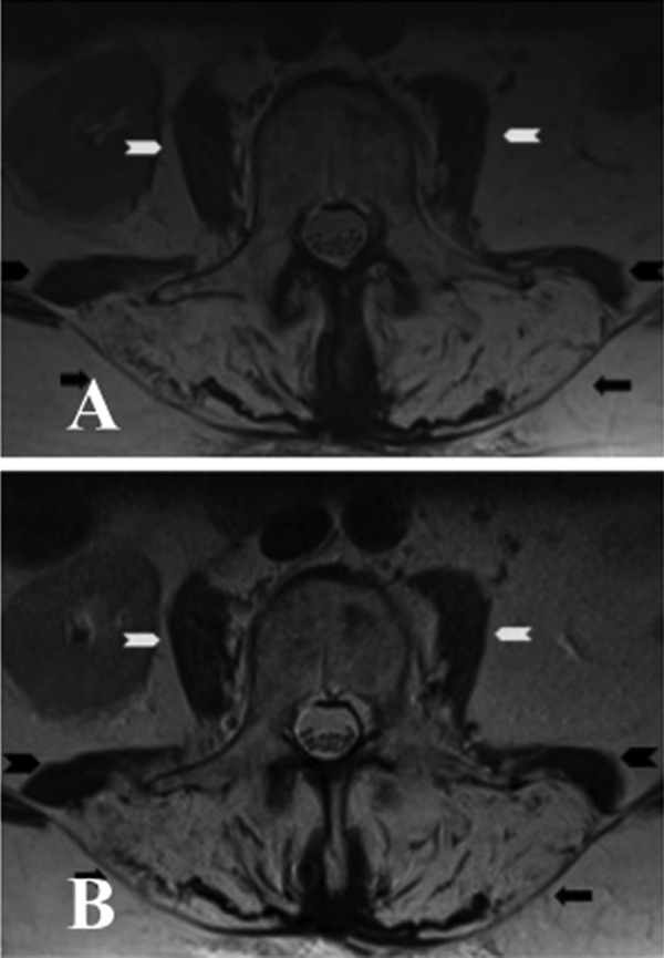
(A) Axial T2 image of the lumbar spine showed marked atrophy of paraspinal muscles (black arrow) bilaterally with increased T2 signals in keeping with fatty replacement. There was also moderate atrophy of psoas (white arrow head) and quadratus lumborum (black arrow head) on both sides. (B) Axial T2 image of the same patient performed 6 months after the initial MRI confirmed persistent fatty atrophy of the paraspinal muscles (black arrow) bilaterally. There was a further very mild increase of atrophy of quadratus lumborum (black arrow head) on both sides. Both psoas (white arrow) did not show any significant change.
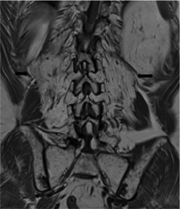
Coronal T1 image showed marked atrophy of paraspinal muscles (black arrow) bilaterally with increased T1 signal in keeping with fatty replacement.
References
-
- Finsterer J, Strobl W. Presentation, etiology, diagnosis, and management of camptocormia. Eur Neurol 2010;2013:1–8 - PubMed
-
- Psimaras D, Maisonobe T, Delanian S, et al. Late onset radiation-induced camptocormia. J Neurol 2011;2013:1723–5 - PubMed
-
- Furby A, Behin A, Lefaucheur JP, et al. Late-onset cervicoscapular muscle atrophy and weakness after radiotherapy for Hodgkin disease: a case series. J Neurol Neurosurg Psychiatry 2010;2013:101–4 - PubMed
-
- Bowen J, Gregory R, Squier M, et al. The post-irradiation lower motor neuron syndrome neuronopathy or radiculopathy? Brain 1996;2013(Pt 5):1429–39 - PubMed
-
- Carver BS, Al-Ahmadie H, Sheinfeld J. Adult and pediatric testicular teratoma. Urol clin North Am 2007;2013:245–51 - PubMed
Publication types
MeSH terms
Supplementary concepts
LinkOut - more resources
Full Text Sources
Other Literature Sources
Medical
Molecular Biology Databases