

Surgical management of scalp arterio-venous malformation and scalp venous malformation: An experience of eleven cases
- PMID: 23960313
- PMCID: PMC3745130
- DOI: 10.4103/0970-0358.113723
Surgical management of scalp arterio-venous malformation and scalp venous malformation: An experience of eleven cases
Abstract
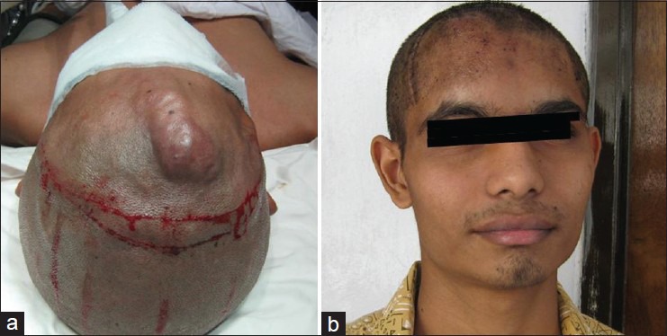
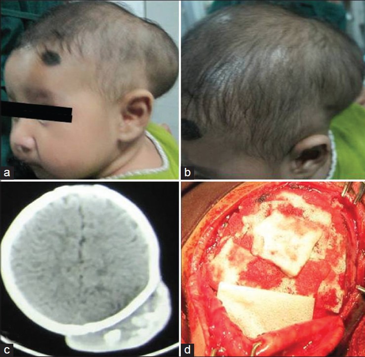
Aims: Scalp arterio-venous malformation (AVM) and scalp venous malformation (SVM) are rare conditions that usually need surgical treatment. Here, we have reported our experience of the surgical management of such lesions with a short review of the literature.
Materials and methods: In this prospective study, 11 patients with scalp AVM and SVM, who underwent surgical excision of lesion in our hospital from 2006 to 2012, were included. All suspected high-flow AVM were investigated with the selective internal and external carotid digital subtraction angiogram (DSA) ± computed tomography (CT) scan of brain with CT angiogram or magnetic resonance imaging (MRI) of brain with MR angiogram, and all suspected low-flow vascular malformation (VM) was investigated with MRI of brain + MR angiogram. Eight were high-flow and three were low-flow VM.
Results: All lesions were successfully excised. Scalp cosmetic aspects were acceptable in all cases. There was no major post-operative complication or recurrence till last follow-up.
Conclusions: With preoperative appropriate surgical planning, scalp AVM and SVM can be excised without major complication.
Keywords: Arterio-venous malformation; cavernous angioma; cirsoid aneurysm; scalp AVM; scalp venous malformatiom; sinus pericranii.
Conflict of interest statement
Figures


References
-
- Senoglu M, Yasim A, Gokce M, Senoglu N. Nontraumatic scalp arteriovenous fistula in an adult: Technical report on an illustrative case. Surg Neurol. 2008;70:194–7. - PubMed
-
- Muthukumar N, Rajagopal V, Manoharan AV, Durairaj N. Surgical management of cirsoid aneurysms. Acta Neurochir (Wien) 2002;144:349–56. - PubMed
-
- Shenoy SN, Raja A. Scalp arteriovenous malformations. Neurol India. 2004;52:478–81. - PubMed
-
- Massimi L, De Bonis P, Esposito G, Novegno F, Pettorini B, Tamburrini G, et al. Vertex scalp mass as presenting sign of a complex intracranial vascular malformation. J Neurosurg Pediatr. 2009;3:307–10. - PubMed
LinkOut - more resources
Full Text Sources
Other Literature Sources