

Assessment of cardiac involvement of hepatitis C virus; tissue Doppler imaging and NTproBNP study
- PMID: 23960652
- PMCID: PMC3727462
- DOI: 10.1016/j.jsha.2011.04.005
Assessment of cardiac involvement of hepatitis C virus; tissue Doppler imaging and NTproBNP study
Abstract
Introduction: Hepatitis C disease burden is substantially increasing in Egyptian community, it is estimated that prevalence of Hepatitis C virus (HCV) in Egyptian community reach 22% of total population. Recently there is a global alert of HCV cardiovascular complications.
Objective: To evaluate LV diastolic functions of HCV patients using tissue Doppler Imaging and NTPBNP.
Methods: 30 HCV patients of 30 years, sex & BMI matched controls were evaluated by PCR, ECG, Echocardiography "conventional Doppler, pulsed wave tissue Doppler (PW-TD), strain rate imaging" & NTPBNP to assess LV diastolic functions. Mean age was 32.8 years ± 5.1 in HCV group, 29.8 years ± 6.6 in control group. Cardiovascular anomalies and predisposing factors were excluded.
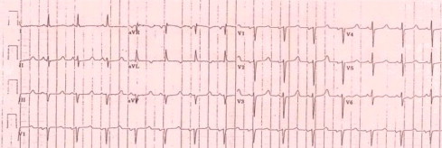
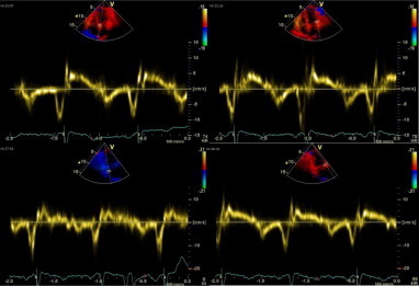
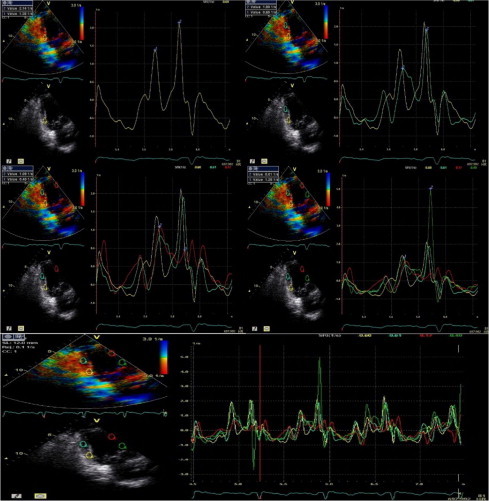
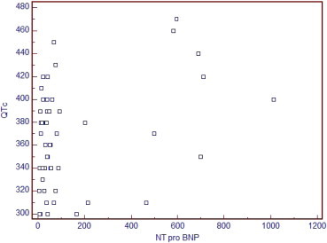
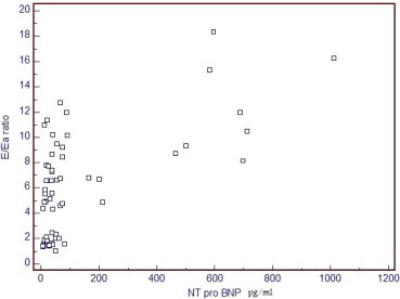
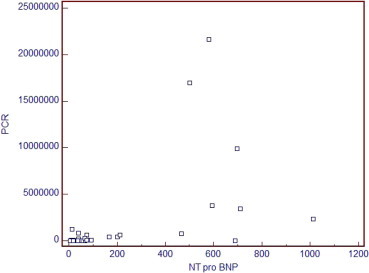
Results: HCV group has shown significant increase in QTc interval, significant statistical increase in A wave, deceleration time; (p < 0.05), highly significant decrease in tissue Doppler E a (p < 0.001), highly significant decrease in A a (p < 0.001), highly significant increased E/E a ratio (p value < 0.001), significant decrease in E a/A a ratio and significant increase in SRa (p < 0.05). NTPBNP levels showed highly significant increase with mean value 222 pg/ml ± 283 in HCV group and 32.7 pg/ml ± 21.2 in control group (p value < 0.001). The best cut-off value of NTPBNP to detect diastolic dysfunction in HCV group was 213 pg/ml. No statistical differences in SRe/SRa and E/SRe ratios were observed, however they had significant correlation with NTPBNP level and tissue Doppler parameters. The best cut-off value of E/SRe ratio to detect diastolic dysfunction in HCV group was 0.91, with 75% sensitivity and 100% specificity.
Conclusion and recommendation: This data show the first direct evidence that HCV infection causes diastolic dysfunction without any other predisposing factors, probably due to chronic inflammatory reaction with mild fibrosis in the heart. Previous studies did not follow strict inclusion and exclusion criteria that confirm the independent role of HCV to cause diastolic dysfunction. Tissue Doppler was more sensitive to diagnose diastolic dysfunction than conventional Doppler.
Keywords: Cardiomyopathy; Diastolic dysfunction; Hepatitis C virus; NTproBNP; Strain rate imaging; Tissue Doppler imaging.
Figures
References
-
- Antonelli A. High levels of circulating N-terminal pro-brain natriuretic peptide in patients with hepatitis C. J. Viral Hepat. 2010;17(12):851–853. - PubMed
-
- Asbjørn Støylen, 2005. Strain rate imaging. Cardiac deformation imaging by ultrasound/echocardiography-tissue Doppler and Speckle tracking. Retrieved online from <http://folk.ntnu.no/stoylen/strainrate/>.
-
- Deffic-Burban S. Expected increase in hepatitis C-related mortality in Egypt due to pre-2000 infections. J. Hepatol. 2006;44(3):455–461. - PubMed
-
- Demirdal T. Cardiomyopathy in Patients with Chronic Hepatitis C Virus. ICC Munich; Germany: 2007. (17th European Congress of Clinical Microbiology and Infectious Diseases).
-
- Dimitroulas T. Early detection of cardiac involvement in systemic sclerosis assessed by tissue-Doppler echocardiography: relationship with neurohormonal activation and endothelial dysfunction. J. Rheumatol. 2010;37(5):993–999. - PubMed
LinkOut - more resources
Full Text Sources
