

Orbital retinoblastoma: Present status and future challenges - A review
- PMID: 23960917
- PMCID: PMC3729383
- DOI: 10.1016/j.sjopt.2010.10.010
Orbital retinoblastoma: Present status and future challenges - A review
Abstract
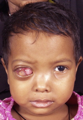
Orbital retinoblastoma is a catastrophic event traditionally carrying a dismal prognosis. Although its incidence is less in the developed countries it continues to be one of the major diagnosis at presentation in the developing world. Orbital retinoblastoma encompasses a wide range of distinct clinical entities with varying tumor load. There are no standard treatment protocols as of now but the current preferred management is multimodal with a combination of initial high-dose chemotherapy, surgery, external beam radiotherapy and prolonged chemotherapy for twelve cycles. In spite of progress on all fronts including surgical, medical, diagnostic, genetic and rehabilitative with improving survival rates, however, lack of access to medical facilities, lack of education about the need for early medical attention and cultural resistance to enucleation continue to contribute to an epidemic of extra ocular disease at diagnosis in the developing world. This review introduces the various terminologies used in the spectrum of orbital retinoblastoma, discusses in details the clinical aspects and management protocols, current status and the future directions.
Keywords: Chemotherapy; Extra ocular; Orbit; Proptosis; Radiotherapy; Retinoblastoma.
Figures


References
-
- Abramson D.H., Niksarli K., Ellsworth R.M. Changing trends in the management of retinoblastoma 1951–1965 vs 1966–1980. J. Pediatr. Ophthalmol. Strabismus. 1994;31:32–37. - PubMed
-
- Abramson D.H., Beaverson K., Sangani P. Screening for retinoblastoma: presenting signs as prognosticators of patient and ocular survival. Paediatrics. 2003;112:1248–1255. - PubMed
-
- Acquaviva A., Barberi L., Bernardini C. Medical therapy of retinoblastoma in children. J. Neurosurg. Sci. 1982;26:49–52. - PubMed
-
- Ajaiyeoba I.A., Akang E.E., Campbell O.B. Retinoblastomas in Ibadan: treatment and prognosis. West Africa J. Med. 1993;12:223–227. - PubMed
-
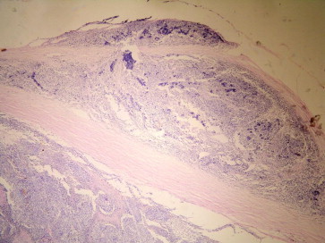
- Ali, M.J., Gupta, R., Vemuganti, G.K., Honavar, S.G., 2009. Histopathology of retinoblastoma after primary chemotherapy. In: Proceedings of the XIV International Congress of Ocular Oncology, Cambridge, p. 296.
LinkOut - more resources
Full Text Sources
