

Value of positron emission tomography/computed tomography in diagnosis and staging of primary ocular and orbital tumors
- PMID: 23961021
- PMCID: PMC3729316
- DOI: 10.1016/j.sjopt.2012.08.008
Value of positron emission tomography/computed tomography in diagnosis and staging of primary ocular and orbital tumors
Abstract
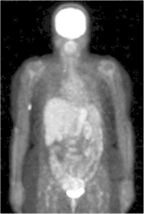
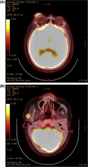
Accurate and reliable staging methods are crucial for optimal care of patients with ocular and orbital malignancies. Positron emission tomography/computed tomography (PET/CT) has recently emerged as a staging tool in the field of ophthalmic oncology. For detecting primary ocular or orbital lesions, PET/CT does not seem to provide an advantage over clinical ophthalmologic examination or conventional imaging studies such as CT or magnetic resonance imaging of the orbit. However, PET/CT may detect distant metastatic lesions that conventional imaging studies miss. For orbital and ocular adnexal lymphoma, use of PET/CT has been proven to be feasible and is now accepted both as a standard part of the initial staging work-up and for the assessment of response to therapy. For other ophthalmic tumors, PET/CT seems most appropriate for advanced metastatic tumors of the orbit, eyelid, and eye, for which the detection of distant metastasis with 1 comprehensive study may be preferable to performing multiple CT scans with contrast.
Keywords: Cancer staging; MALT lymphoma; Orbital lymphoma; Orbital tumors; Positron emission tomography/computed tomography; Uveal melanoma.
Figures
Similar articles
-
Positron emission tomography in the detection and staging of ocular adnexal lymphoproliferative disease.Ophthalmology. 2006 Dec;113(12):2331-7. doi: 10.1016/j.ophtha.2006.05.059. Epub 2006 Sep 25. Ophthalmology. 2006. PMID: 16996604
-
Whole Body Positron Emission Tomography/Computed Tomography (PET/CT) in the Evaluation of Ophthalmic Tumors.Curr Med Sci. 2018 Apr;38(2):310-317. doi: 10.1007/s11596-018-1880-7. Epub 2018 Apr 30. Curr Med Sci. 2018. PMID: 30074190
-
Whole-body 18F-fluorodeoxyglucose positron emission tomography-computed tomography (18F-FDG PET/CT) for staging locally advanced breast cancer: A prospective study from a tertiary cancer centre in south India.Indian J Med Res. 2018 Mar;147(3):256-262. doi: 10.4103/ijmr.IJMR_1368_16. Indian J Med Res. 2018. PMID: 29923514 Free PMC article.
-
A two-way comparison of whole-body 18FDG PET-CT and whole-body contrast-enhanced MRI for distant metastasis staging in patients with malignant tumors: a meta-analysis of 13 prospective studies.Ann Palliat Med. 2020 Mar;9(2):247-255. doi: 10.21037/apm.2020.02.30. Epub 2020 Mar 18. Ann Palliat Med. 2020. PMID: 32233618 Review.
-
PET/CT and PET/MRI in ophthalmic oncology (Review).Int J Oncol. 2020 Feb;56(2):417-429. doi: 10.3892/ijo.2020.4955. Epub 2020 Jan 3. Int J Oncol. 2020. PMID: 31939615 Free PMC article. Review.
Cited by
-
Orbital Metastases: When to Suspect? When to biopsy?Middle East Afr J Ophthalmol. 2018 Apr-Jun;25(2):60-64. doi: 10.4103/meajo.MEAJO_93_18. Middle East Afr J Ophthalmol. 2018. PMID: 30122850 Free PMC article. Review.
-
Advances in Positron Emission Tomography/Computed Tomography for Diagnosing and Managing Primary Posterior Uveal Melanoma.Med Sci Monit. 2025 Aug 19;31:e949252. doi: 10.12659/MSM.949252. Med Sci Monit. 2025. PMID: 40826825 Free PMC article. Review.
-
Conjunctival Lymphoma.Eye (Lond). 2023 Apr;37(5):837-848. doi: 10.1038/s41433-022-02176-2. Epub 2022 Jul 26. Eye (Lond). 2023. PMID: 35882984 Free PMC article. Review.
-
The practice patterns in the management of sebaceous carcinoma of the eyelid in the Asia Pacific region.Eye (Lond). 2019 Sep;33(9):1433-1442. doi: 10.1038/s41433-019-0432-0. Epub 2019 Apr 5. Eye (Lond). 2019. PMID: 30952958 Free PMC article.
-
Update in oculoplastic imaging.Saudi J Ophthalmol. 2012 Oct;26(4):347-8. doi: 10.1016/j.sjopt.2012.11.001. Epub 2012 Nov 10. Saudi J Ophthalmol. 2012. PMID: 23961018 Free PMC article. No abstract available.
References
-
- Kwee T.C., Kwee R.M., Nievelstein R.A. Imaging in staging of malignant lymphoma: a systematic review. Blood. 2008;111(2):504–516. - PubMed
-
- Chowdhury F.U., Shah N., Scarsbrook A.F., Bradley K.M. [18F]FDG PET/CT imaging of colorectal cancer: a pictorial review. Postgrad Med J. 2010;86(1013):174–182. - PubMed
-
- Schoder H., Larson S.M., Yeung H.W. PET/CT in oncology: integration into clinical management of lymphoma, melanoma, and gastrointestinal malignancies. J Nucl Med. 2004;45(Suppl. 1):72S–81S. - PubMed
-
- von Schulthess G.K., Steinert H.C., Hany T.F. Integrated PET/CT: current applications and future directions. Radiology. 2006;238(2):405–422. - PubMed
-
- Messa C., Bettinardi V., Picchio M., Pelosi E., Landoni C., Gianolli L. PET/CT in diagnostic oncology. Q J Nucl Med Mol Imaging. 2004;48(2):66–75. - PubMed
LinkOut - more resources
Full Text Sources
