

Glaucoma masqueraders: Diagnosis by spectral domain optical coherence tomography
- PMID: 23961029
- PMCID: PMC3729362
- DOI: 10.1016/j.sjopt.2012.08.006
Glaucoma masqueraders: Diagnosis by spectral domain optical coherence tomography
Abstract
Background: Advances in optic nerve and retinal imaging have dramatically changed the care of glaucoma patients, complementing the importance of the clinical exam of the optic nerve and automated perimetry in making the diagnosis of glaucoma. Computerized imaging, however, does not replace the clinical exam, as there can be overlap in the appearance of non-glaucomatous optic neuropathies with glaucoma.
Methods: The spectral domain optic coherence tomography (SD-OCT) images of five patients with non-glaucomatous optic nerve pathology are presented.
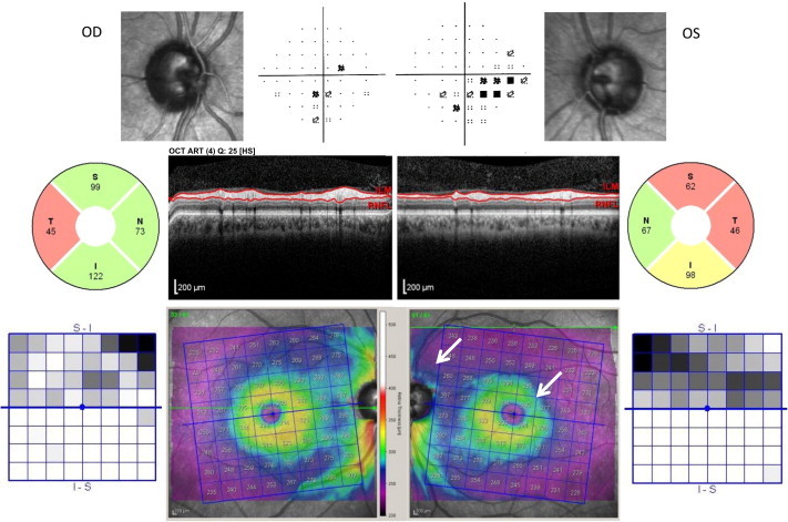
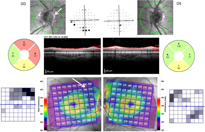
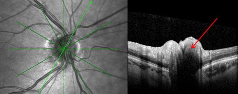
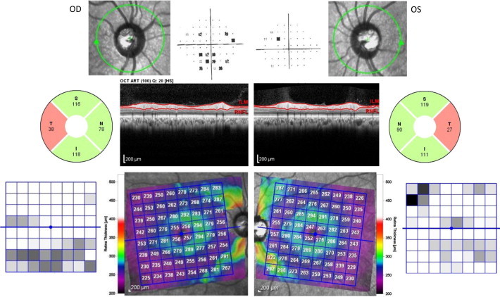
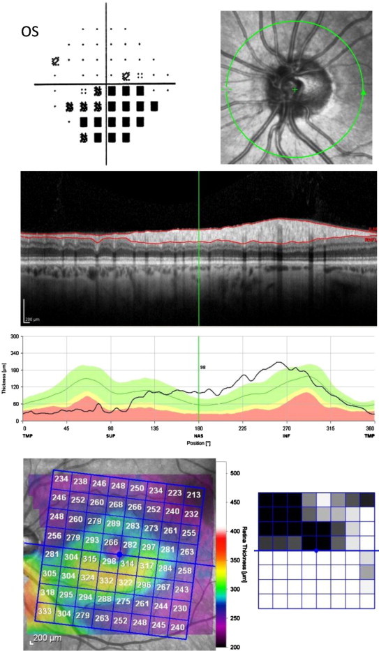
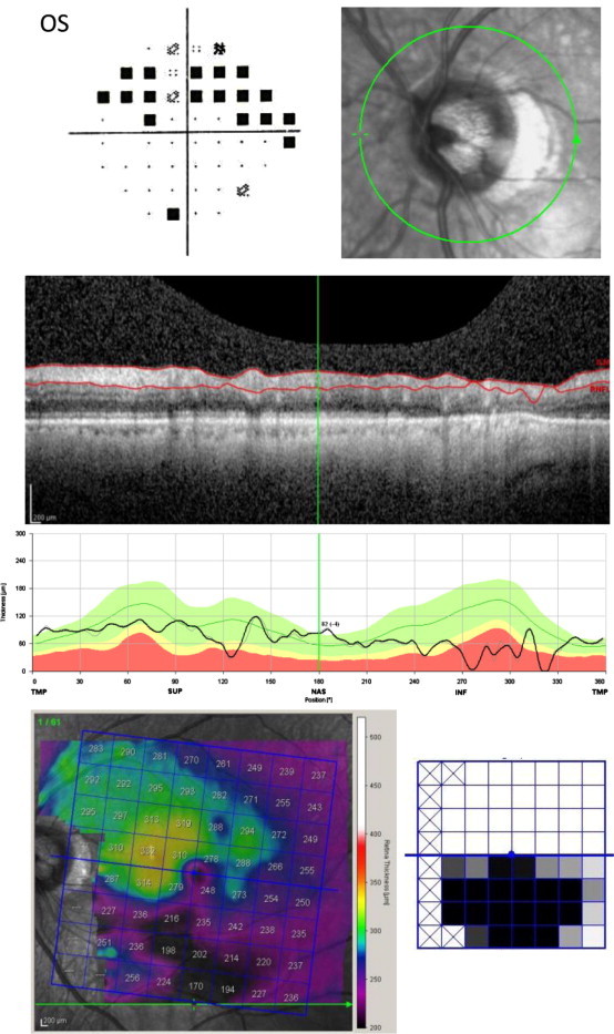
Cases: The first patient had bilateral temporal thinning on OCT imaging and subsequent positive syphilis testing. The second patient had a glaucomatous-appearing inferior arcuate scotoma and associated superior thinning on OCT; these findings were due to buried optic nerve head drusen, clearly appreciated on OCT of the optic nerve head. Bilateral diffuse macular thinning, with preservation of the superior and inferior fiber bundles, was seen in the third patient, who had multiple sclerosis, with no clinical history of optic neuritis. Dense and marked thinning of a macular half, respecting the horizontal meridian, is seen in two patients, one patient with non-arteritic anterior ischemic optic neuropathy and lastly, in a patient with hemi-retinal vein occlusion.
Conclusion: SD-OCT of the optic nerve and retina complements the essential clinical examination of patients with glaucomatous and non-glaucomatous optic neuropathies.
Keywords: Glaucoma; Macular OCT; OCT; Optic neuropathy.
Figures

Similar articles
-
Normal-Tension Glaucoma Masqueraders: Detection Using Optical Coherence Tomography.J Glaucoma. 2017 Apr;26(4):e153-e156. doi: 10.1097/IJG.0000000000000578. J Glaucoma. 2017. PMID: 28121717
-
Retinal nerve fiber layer evaluation in multiple sclerosis with spectral domain optical coherence tomography.Clin Ophthalmol. 2010 Sep 20;4:1007-13. doi: 10.2147/opth.s13278. Clin Ophthalmol. 2010. PMID: 20922034 Free PMC article.
-
Spectral-Domain OCT: Helping the Clinician Diagnose Glaucoma: A Report by the American Academy of Ophthalmology.Ophthalmology. 2018 Nov;125(11):1817-1827. doi: 10.1016/j.ophtha.2018.05.008. Epub 2018 Jul 7. Ophthalmology. 2018. PMID: 30322450
-
[New insights into the study of optic nerve diseases].Nippon Ganka Gakkai Zasshi. 2013 Mar;117(3):187-210; discussion 211. Nippon Ganka Gakkai Zasshi. 2013. PMID: 23631254 Review. Japanese.
-
OCT in the Differential Diagnosis of Optic Neuropathies. A Review.Cesk Slov Oftalmol. 2025;81(2):51-59. doi: 10.31348/2025/15. Cesk Slov Oftalmol. 2025. PMID: 40135703 Review. English.
Cited by
-
Ocular Syphilis Presenting As Non-arteritic Anterior Ischemic Optic Neuropathy.Cureus. 2021 Jul 28;13(7):e16694. doi: 10.7759/cureus.16694. eCollection 2021 Jul. Cureus. 2021. PMID: 34466324 Free PMC article.
-
Neuro-Ophthalmological Optic Nerve Cupping: An Overview.Eye Brain. 2021 Dec 14;13:255-268. doi: 10.2147/EB.S272343. eCollection 2021. Eye Brain. 2021. PMID: 34934377 Free PMC article. Review.
-
Visual field defects and retinal nerve fiber layer damage in buried optic disc drusen: a new insight.Int J Ophthalmol. 2022 Oct 18;15(10):1641-1649. doi: 10.18240/ijo.2022.10.12. eCollection 2022. Int J Ophthalmol. 2022. PMID: 36262850 Free PMC article.
-
Macular thickness analysis for glaucoma diagnosis and management.Taiwan J Ophthalmol. 2016 Jan-Mar;6(1):3-7. doi: 10.1016/j.tjo.2016.01.003. Epub 2016 Feb 28. Taiwan J Ophthalmol. 2016. PMID: 29018702 Free PMC article. Review.
-
A Case of Bilateral Optic Nerve Head Drusen-Induced Inferior Altitudinal Hemianopsia.Neuroophthalmology. 2015 Jun 23;39(4):201-206. doi: 10.3109/01658107.2015.1022899. eCollection 2015 Aug. Neuroophthalmology. 2015. PMID: 27928357 Free PMC article.
References
-
- Savini G., Carbonelli M., Barboni P. Spectral-domain optical coherence tomography for the diagnosis and follow-up of glaucoma. Curr Opin Ophthalmo. 2011;22(2):115–123. - PubMed
-
- Asrani S., Rosdahl J.A., Allingham R.R. Novel software strategy for glaucoma diagnosis: asymmetry analysis of retinal thickness. Arch Ophthalmol. 2011;129(9):1205–1211. - PubMed
-
- Moura F.C. Optical coherence tomography evaluation of retinal nerve fiber layer in longitudinally extensive transverse myelitis. Arq Neuropsiquiatr. 2011;69(1):69–73. - PubMed
-
- Flores-Rodriguez P., Gili P., Martin-Rios M.D. Sensitivity and specificity of time-domain and spectral-domain optical coherence tomography in differentiating optic nerve head drusen and optic disc oedema. Ophthalmic Physiol Opt. 2012;32(3):213–221. - PubMed
LinkOut - more resources
Full Text Sources
