. 2013 Sep 17;52(37):6515-24.
doi: 10.1021/bi400542z. Epub 2013 Sep 6.

Surface reengineering of RPA70N enables cocrystallization with an inhibitor of the replication protein A interaction motif of ATR interacting protein

Affiliations

Surface reengineering of RPA70N enables cocrystallization with an inhibitor of the replication protein A interaction motif of ATR interacting protein

Michael D Feldkamp et al. Biochemistry. .

Abstract

Replication protein A (RPA) is the primary single-stranded DNA (ssDNA) binding protein in eukaryotes. The N-terminal domain of the RPA70 subunit (RPA70N) interacts via a basic cleft with a wide range of DNA processing proteins, including several that regulate DNA damage response and repair. Small molecule inhibitors that disrupt these protein-protein interactions are therefore of interest as chemical probes of these critical DNA processing pathways and as inhibitors to counter the upregulation of DNA damage response and repair associated with treatment of cancer patients with radiation or DNA-damaging agents. Determination of three-dimensional structures of protein-ligand complexes is an important step for elaboration of small molecule inhibitors. However, although crystal structures of free RPA70N and an RPA70N-peptide fusion construct have been reported, RPA70N-inhibitor complexes have been recalcitrant to crystallization. Analysis of the P61 lattice of RPA70N crystals led us to hypothesize that the ligand-binding surface was occluded. Surface reengineering to alter key crystal lattice contacts led to the design of RPA70N E7R, E100R, and E7R/E100R mutants. These mutants crystallized in a P212121 lattice that clearly had significant solvent channels open to the critical basic cleft. Analysis of X-ray crystal structures, target peptide binding affinities, and (15)N-(1)H heteronuclear single-quantum coherence nuclear magnetic resonance spectra showed that the mutations do not result in perturbations of the RPA70N ligand-binding surface. The success of the design was demonstrated by determining the structure of RPA70N E7R soaked with a ligand discovered in a previously reported molecular fragment screen. A fluorescence anisotropy competition binding assay revealed this compound can inhibit the interaction of RPA70N with the peptide binding motif from the DNA damage response protein ATRIP. The implications of the results are discussed in the context of ongoing efforts to design RPA70N inhibitors.


Figures

Figure 1
Figure 1. Subunit and domain structure of RPA
OB-fold domains are depicted as rectangles, the winged helix-turn-helix domain as an octagon and the unstructured phosphorylation domain as an oval. The ssDNA binding domains are RPA70A 70B, 70C and 32D (blue). Domains RPA70N and RPA32C are the primary protein recruitment modules (pink). The RPA trimer is formed by interactions between RPA70C, RPA32D, and RPA14.
Figure 2
Figure 2. Packing of RPA70N in hexagonal and orthorhombic crystal lattices
A) Lattice contacts in the hexagonal crystal lattice between residues E7 and E100 (red) and basic cleft residues R31, R41, and R43 (blue) from an adjacent RPA70N molecule. B) Packing of the basic cleft of RPA70N against the backside of its symmetry mate in the hexagonal crystal lattice blocks access to the basic cleft (dashed line). C) Packing in the orthorhombic lattice adopted by RPA70N charge reversal mutants produces solvent channels formed directly by the basic clefts (dashed line) of opposing RPA70N symmetry mates.
Figure 3
Figure 3. Comparison of the structures of WT RPA70N and the E7R, E100R, and E7R, E100R mutants
A) Best-fit superposition (Cα atoms) of the structures of E7R (red), E100R (blue), and E7R-E100R (cyan) on the structure of WT RPA70N (green).
Figure 4
Figure 4. Electrostatic surface potentials of WT RPA70N and the E7R, E100R, and E7R, E100R mutants
Surface potentials calculated by the APBS software for (from left to right) WT, E7R, E100R and E7R, E100R. Dashed circles are drawn to highlight the basic cleft, residue 7, and residue 100.
Figure 5
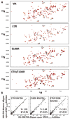
Figure 5. Comparison of NMR chemical shift perturbations induced by binding of ATRIP54-68 in WT RPA70N and the E7R, E100R, and E7R, E100 mutants
A) NMR titration of WT RPA70N and the E7R, E100R, and E7R, E100 mutants with ATRIP54-68. Overlay of 15N-1H HSQC spectra of WT RPA70N and the E7R, E100R, and E7R, E100R mutants as ATRIP54-68 is titrated into the sample. Base 15N-1H crosspeaks transition from yellow to dark red upon ATRIP54-68 saturation. Arrows indicate the starting and ending points for select residues exhibiting chemical shift perturbations as ATRIP54-68 is added. B) Correlation plots comparing the chemical shifts in complexes with ATRIP54-68 for mutant (y-axis) and WT RPA70N (x-axis).
Figure 6
Figure 6. Structure of RPA70N E7R in complex with VU079104
A) Chemical structure of VU079104. B) Structure of RPA70N E7R colored to highlight side chains (cyan) within 3.5 Å of VU079104 (green), a crystallographic water (red sphere), and hydrogen bonding interactions to VU079104 (yellow dashes). VU079104 2Fo-Fc and Fo-Fc electron density maps are displayed contoured at 1σ and 3 σ in blue and red mesh respectively. C) Changes in E7R side chains induced by binding of the ligand. The overlay shows the positioning of side chains within 3.5 Å of VU079104 (green) for E7R (purple sticks) and the E7R-VU079104 complex (cyan). D) Superposition of VU079104 (sticks) from the structure of the complex onto the surface of the structure of the RPA70N-p53 complex (PDB ID: 2B3G). Solvent exposed surface residues within 4 Å of VU079104 and p53 peptide (black) are colored yellow and red, respectively.
Figure 7
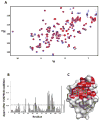
Figure 7. 15N-1H chemical shift changes in WT-RPA70N induced by binding of VU079104
A) Overlaid 15N-1H spectra of WT-RPA70N (blue) and VU079104 saturated WT-RPA70N (red), arrows indicated the direction of movement of select 15N-1H cross peaks. B) Quantified chemical shift perturbations induced upon binding of VU079104 to WT RPA70N. C) Locations of significantly perturbed Wt-RPA70N residues (red) upon VU079104 addition mapped upon the structure of E7R RPA70N in complex with VU079014 (green sticks).
Figure 8
Figure 8. Fluorescence polarization anisotropy assay of the displacement of ATRIP54-68 from RPA70N by VU079104
Concentration–response curve used for determination of the Kd for binding of VU079104 to WT-RPA70N. The error bars represent standard deviations of two independent experiments, each performed in duplicate. The data point represented by the square symbol corresponds to the anisotropy value recorded for ATRIP54-68 in the absence of RPA70N.
Figure 9
Figure 9. Comparative structural analysis suggests targeting of the S55 pocket to increase the affinity of the VU079104 framework for RPA70N
Superposition of VU079104 onto the surface of the structure of the RPA70N-p53 peptide fusion (PDBID: 2B3G) showing the close proximity of VU079104 to the hydrophobic pocket into which the F54 ring of p53 is inserted (red), and a basic patch on RPA70N created by residues R43 and R91 (blue).

References

    1. Wold MS, Kelly T. Purification and characterization of replication protein A, a cellular protein required for in vitro replication of simian virus 40 DNA. Proc Natl Acad Sci U S A. 1988;85:2523–2527. - PMC - PubMed
    1. Wold MS. Replication protein A: a heterotrimeric, single-stranded DNA-binding protein required for eukaryotic DNA metabolism. Annu Rev Biochem. 1997;66:61–92. - PubMed
    1. Bochkarev A, Pfuetzner RA, Edwards AM, Frappier L. Structure of the single-stranded-DNA-binding domain of replication protein A bound to DNA. Nature. 1997;385:176–181. - PubMed
    1. Lao Y, Gomes XV, Ren Y, Taylor JS, Wold MS. Replication protein A interactions with DNA. III. Molecular basis of recognition of damaged DNA. Biochemistry. 2000;39:850–859. - PubMed
    1. Treuner K, Ramsperger U, Knippers R. Replication protein A induces the unwinding of long double-stranded DNA regions. J Mol Biol. 1996;259:104–112. - PubMed

Publication types

MeSH terms

Substances