. 2013 Sep 18;135(37):13710-9.
doi: 10.1021/ja400146z. Epub 2013 Sep 6.

Influenza virus A M2 protein generates negative Gaussian membrane curvature necessary for budding and scission

Affiliations

Influenza virus A M2 protein generates negative Gaussian membrane curvature necessary for budding and scission

Nathan W Schmidt et al. J Am Chem Soc. .

Abstract

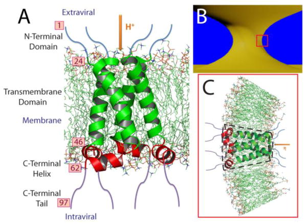
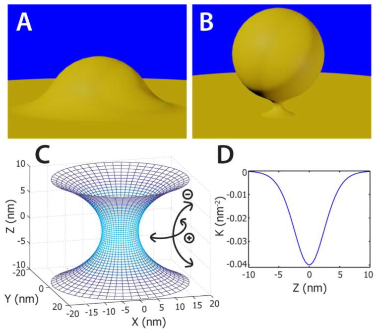
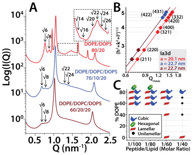
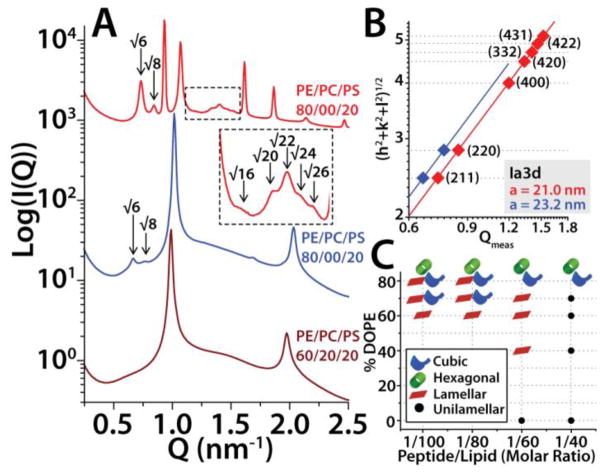
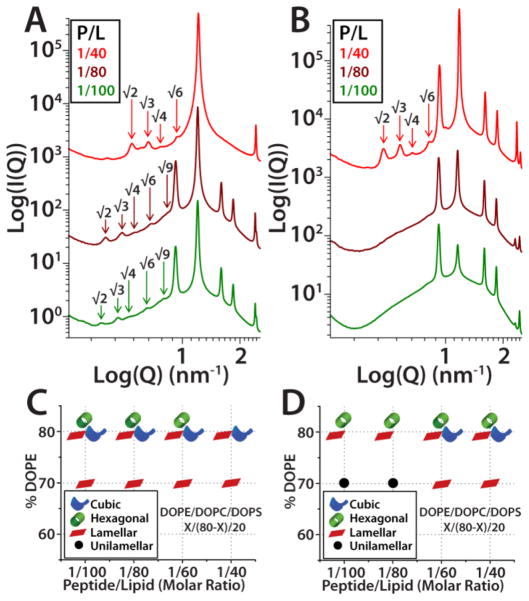
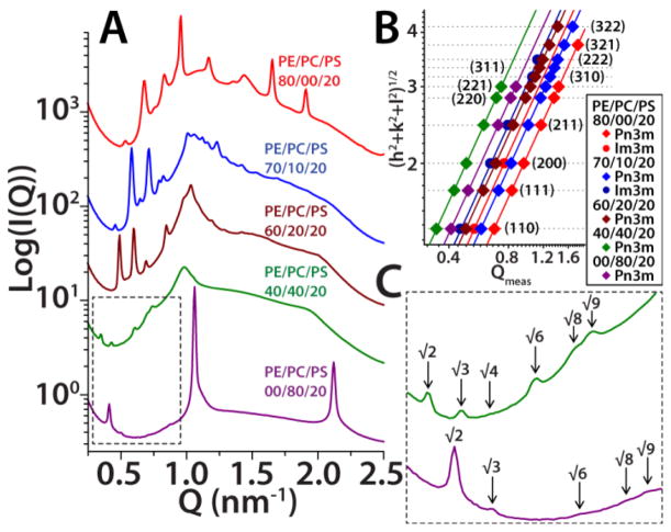
The M2 protein is a multifunctional protein, which plays several roles in the replication cycle of the influenza A virus. Here we focus on its ability to promote budding of the mature virus from the cell surface. Using high-resolution small-angle X-ray scattering we show that M2 can restructure lipid membranes into bicontinuous cubic phases which are rich in negative Gaussian curvature (NGC). The active generation of negative Gaussian membrane curvature by M2 is essential to influenza virus budding. M2 has been observed to colocalize with the region of high NGC at the neck of a bud. The structural requirements for scission are even more stringent than those for budding, as the neck must be considerably smaller than the virus during 'pinch off'. Consistent with this, the amount of NGC in the induced cubic phases suggests that M2 proteins can generate high curvatures comparable to those on a neck with size 10× smaller than a spherical influenza virus. Similar experiments on variant proteins containing different M2 domains show that the cytoplasmic amphipathic helix is necessary and sufficient for NGC generation. Mutations to the helix which reduce its amphiphilicity and are known to diminish budding attenuated NGC generation. An M2 construct comprising the membrane interactive domains, the transmembrane helix and the cytoplasmic helix, displayed enhanced ability to generate NGC, suggesting that other domains cooperatively promote membrane curvature. These studies establish the importance of M2-induced NGC during budding and suggest that antagonizing this curvature is a viable anti-influenza strategy.


Figures

Figure 1
Figure 1
The influenza A virus M2 protein has an essential role in viral budding and scission. A. Domain Structure model of influenza A virus M2 proton channel. Model is based on ref . B. Illustration of the neck in a bud. C. Close up view illustrating an M2 channel on the neck region corresponding to the red box in B. M2 generates membrane curvature via its C-terminal amphipathic helix, as well as from the volume excluded by the membrane-associated regions of the channel (outlined by dashed lines).
Figure 2
Figure 2
Influenza A viral budding and scission is mediated by the M2 protein. A. Illustration of an early stage in the budding process when the emerging virion begins to protrude out of the cytoplasmic membrane. The M2 protein localizes to the base of the protrusion which has negative Gaussian curvature (positive curvature around the bump and initial negative curvature up the bump). As the bud progresses the virus emerges as the base constricts into a neck with higher NGC. B. Late stage in the budding process just before membrane scission. The virus is approximated as a sphere with a diameter that is 10x the size of the width of the constricted neck. C. A catenoid surface with 10 nm width at its narrowest cross-section, Z = 0 nm. The shape of the neck will conform to a catenoid, a minimal surface which has zero mean curvature and NGC everywhere. Arrows show directions of positive and negative curvature. D. Negative Gaussian curvature, K, along the vertical direction of the neck in C. The K values on the surface of this catenoid are similar to those extracted from the bicontinuous cubic phases that M2 proteins generated in membranes.
Figure 3
Figure 3
The M2 protein generated NGC in phospholipid membranes. A. SAXS spectra from DOPE/DOPC/DOPS = X/(80−X)/20 vesicles incubated with M2 at 1/100 protein to lipid (P/L) molar ratio. M2 induced Ia3d cubic phases in membranes with DOPE content as low as X = 60%. Overall, negative intrinsic curvature lipids promoted non-lamellar phase formation as indicated by the appearance of higher reflections from the Ia3d at elevated DOPE, and the presence of an inverted hexagonal phase in 80% DOPE membranes. The inset provides a more detailed view of the higher order Ia3d phase reflections. B. Log-Log plots of the assigned reflections versus the measured Q-positions for Ia3d cubic peaks in the three spectra from A. For powder averaged cubic phases Q(hkl) = 2π√(h2+k2+l2)/a, where h, k, & l, are the Miller indices and a is the lattice parameter. The calculated lattice parameters, extracted using linear trendline fits, are the same color as the spectra in A. Left-shifted trendlines indicate larger cubic phase lattice parameters. C. Phase diagram for M2 protein with ternary DOPE/DOPC/DOPS membranes. The symbols show which phases were observed for a given %DOPE and P/L ratio. In PE-rich membranes non-lamellar cubic and inverted hexagonal phases were prominent, whereas lamellar phases were dominant in reduced DOPE membranes.
Figure 4
Figure 4
The Ala-M2 protein displayed reduced ability to generate NGC compared with M2. A. Analogous SAXS spectra for Ala-M2 with DOPE/DOPC/DOPS = X/(80−X)/20 membranes at P/L = 1/100. A DOPE concentration of X = 70% is required for Ala-M2 to induce the Ia3d cubic phase. B. Indexation of the Ia3d cubic phases for DOPE/DOPC/DOPS = 80/20/00 (red), and 70/10/20 (blue) membrane samples. The procedure for extracting cubic phase lattice parameters is identical to Figure 3. The cubic phase lattice parameter increased with decreasing membrane DOPE concentration. C. Phase diagram for Ala-M2. Compared to M2, Ala-AM2 showed increased propensity to induce inverted hexagonal phases, and was less able to induce cubic phases.
Figure 5
Figure 5
A peptide consisting of the M2 cytoplasmic amphipathic α-helix (45–62) is sufficient to generate membrane NGC. A. In DOPS/DOPE = 20/80 membranes the M2 C-cyto peptide induced Pn3m cubic phases over a variety of P/L ratios. The shift of the cubic phase reflections to higher Q-values with increasing P/L, shows that increased amounts of peptide generate Pn3m cubics with smaller lattice parameters. B. Compared to the native peptide, the penta-Ala substituted M2 C-cyto showed reduced ability to generate NGC. At P/L = 1/100 & 1/80, Ala-M2 C-cyto induced only lamellar and inverted hexagonal phases; the Pn3m cubic appeared at higher P/L ratios only. C., & D. The phase diagrams for M2 C-cyto, and Ala-M2 C-cyto. The native M2 C-cyto peptide showed better membrane restructuring ability than the penta-Ala variant, and generated NGC over a wider region of the phase diagram.
Figure 6
Figure 6
The amino acid composition of the M2 C-cyto peptide is similar to membrane active antimicrobial peptides. Since arginine can generate both positive and negative curvature while lysine generates negative curvature only and hydrophobicity generates positive curvature only, the requirement that AMPs generate NGC implies that a decrease in peptide arginine content (NR) can be offset by an increase in lysine (NK) plus hydrophobicity. A. & B. show the positive relationship between lysine and average hydrophobicity for 1080 AMPs (gray circles) using the Eisenberg Consensus and Wimley-White Biological hydrophobicity scales, respectively. The M2 C-cyto peptide (red) tends to be more hydrophobic than an average AMP with equivalent Lys to Arg ratio, and the Ala-M2 C-cyto (blue) is less hydrophobic than WT. C. & D. are histograms comparing the distribution of average AMP hydrophobicities (gray bars) with those of C-cyto domain peptides (M2 C-cyto red square, Ala-M2 C-cyto blue square) accounting for only the hydrophobic residues. C uses Eisenberg Consensus scale, and D uses the Wimley-White Biological scale. Both M2-derived peptides lie within the hydrophobic range of AMPs and alanine substitution results in a less hydrophobic C-cyto domain.
Figure 7
Figure 7
The M2TM-cyto peptide generated NGC over the entire range of tested lipid compositions. A. In DOPE/DOPC/DOPS = X/(80−X)/20 membranes, M2TM-cyto induced the coexisting Im3m and Pn3m cubic phases in PE-rich, X ≥ 70% membranes, while at lower concentrations of c0 < 0 lipids the Pn3m phase was observed. In general, at fixed P/L, the reflections from the Pn3m shift to lower Q-values with decreasing membrane PE concentration. P/L = 1/60 for all membrane compositions except PE/PC/PS = 00/80/20 which is at P/L = 1/40. B. Indexation of the Pn3m and Im3m cubic phases in A. A left shift of trendlines indicates cubic phases with larger lattice parameters. C. Close-up view of the Pn3m cubic phase peaks in low-PE membrane spectra from A.
Figure 8
Figure 8
The Ala-M2TM-cyto peptide variant generated NGC over a smaller range of lipid compositions compared with M2TM-cyto. A. At P/L = 1/60, Ala-M2TM-cyto induced coexisting Im3m and Pn3m cubic phases in DOPE/DOPC/DOPS = 80/00/20, & 70/10/20 membranes, and in 60/20/20 membranes induced a pure Pn3m cubic. In membranes with reduced DOPE, only lamellar phases were observed. The phase diagrams for M2TM-cyto, B., and Ala-M2TM-cyto, C., peptides with ternary DOPE/DOPC/DOPS = X/(80−X)/20 lipid compositions. The native M2TM-cyto peptide generated NGC over a wider region of the phase diagram, while the Ala-substituted version displayed greater ability to generate inverted hexagonal phases.

Similar articles

Cited by

References

    1. Chen BJ, Lamb RA. Virology. 2008;372:221. - PMC - PubMed
    1. Carlton JG, Martin-Serrano J. Biochem Soc Trans. 2009;37:195. - PubMed
    1. Pornillos O, Garrus JE, Sundquist WI. Trends in Cell Biol. 2002;12:569. - PubMed
    1. Bruce EA, Medcalf L, Crump CM, Noton SL, Stuart AD, Wise HM, Elton D, Bowers K, Digard P. Virology. 2009;390:268. - PubMed
    1. Rossman JS, Jing X, Leser GP, Lamb RA. Cell. 2010;142:902. - PMC - PubMed

Publication types

MeSH terms