

The chromosome cycle of prokaryotes
- PMID: 23962352
- PMCID: PMC3800152
- DOI: 10.1111/mmi.12372
The chromosome cycle of prokaryotes
Abstract
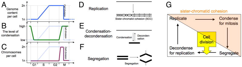
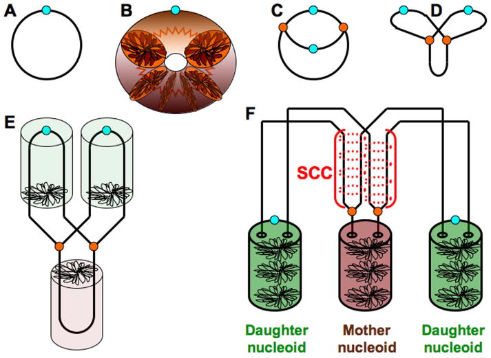
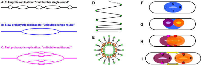
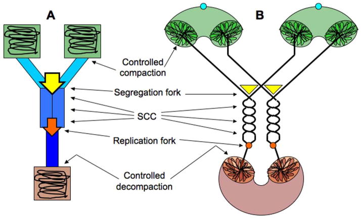
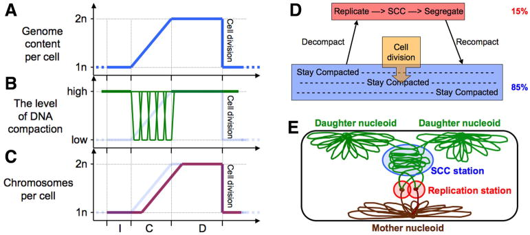
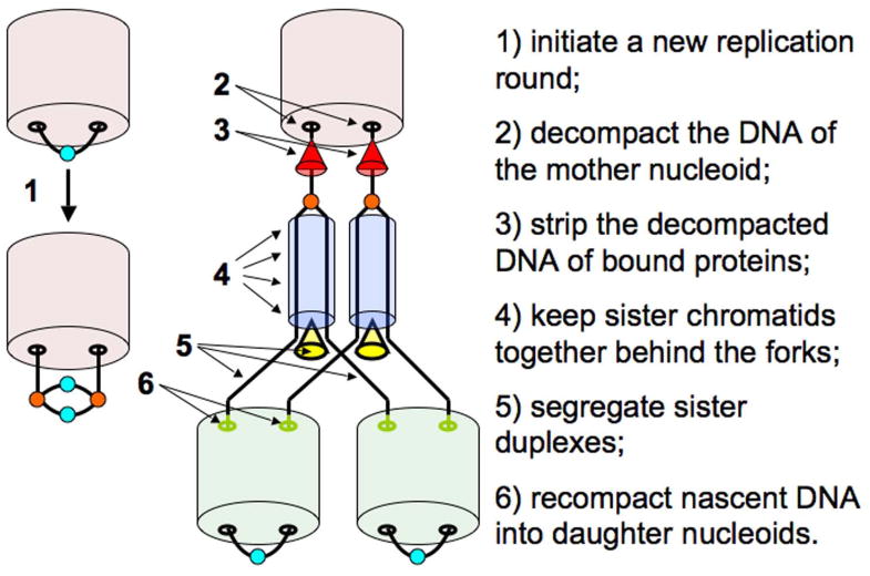
In both eukaryotes and prokaryotes, chromosomal DNA undergoes replication, condensation-decondensation and segregation, sequentially, in some fixed order. Other conditions, like sister-chromatid cohesion (SCC), may span several chromosomal events. One set of these chromosomal transactions within a single cell cycle constitutes the 'chromosome cycle'. For many years it was generally assumed that the prokaryotic chromosome cycle follows major phases of the eukaryotic one: -replication-condensation-segregation-(cell division)-decondensation-, with SCC of unspecified length. Eventually it became evident that, in contrast to the strictly consecutive chromosome cycle of eukaryotes, all stages of the prokaryotic chromosome cycle run concurrently. Thus, prokaryotes practice 'progressive' chromosome segregation separated from replication by a brief SCC, and all three transactions move along the chromosome at the same fast rate. In other words, in addition to replication forks, there are 'segregation forks' in prokaryotic chromosomes. Moreover, the bulk of prokaryotic DNA outside the replication-segregation transition stays compacted. I consider possible origins of this concurrent replication-segregation and outline the 'nucleoid administration' system that organizes the dynamic part of the prokaryotic chromosome cycle.
© 2013 John Wiley & Sons Ltd.
Conflict of interest statement
The author declares no conflict of interest.
Figures
Similar articles
-
The precarious prokaryotic chromosome.J Bacteriol. 2014 May;196(10):1793-806. doi: 10.1128/JB.00022-14. Epub 2014 Mar 14. J Bacteriol. 2014. PMID: 24633873 Free PMC article. Review.
-
Bacterial chromosome dynamics.Science. 2003 Aug 8;301(5634):780-5. doi: 10.1126/science.1084780. Science. 2003. PMID: 12907786 Review.
-
Partitioning of the Escherichia coli chromosome: superhelicity and condensation.Biochimie. 2001 Jan;83(1):41-8. doi: 10.1016/s0300-9084(00)01204-9. Biochimie. 2001. PMID: 11254973 Review.
-
The Origin of Chromosomal Replication Is Asymmetrically Positioned on the Mycobacterial Nucleoid, and the Timing of Its Firing Depends on HupB.J Bacteriol. 2018 Apr 24;200(10):e00044-18. doi: 10.1128/JB.00044-18. Print 2018 May 15. J Bacteriol. 2018. PMID: 29531181 Free PMC article.
-
[The bacterial cell cycle: DNA replication, nucleoid segregation, and cell division].Mikrobiologiia. 2005 Jul-Aug;74(4):437-51. Mikrobiologiia. 2005. PMID: 16211846 Review. Russian.
Cited by
-
Static and Dynamic Factors Limit Chromosomal Replication Complexity in Escherichia coli, Avoiding Dangers of Runaway Overreplication.Genetics. 2016 Mar;202(3):945-60. doi: 10.1534/genetics.115.184697. Epub 2016 Jan 22. Genetics. 2016. PMID: 26801182 Free PMC article.
-
Creating Polyploid Escherichia Coli and Its Application in Efficient L-Threonine Production.Adv Sci (Weinh). 2023 Nov;10(31):e2302417. doi: 10.1002/advs.202302417. Epub 2023 Sep 25. Adv Sci (Weinh). 2023. PMID: 37749873 Free PMC article.
-
Archaeal imaging: leading the hunt for new discoveries.Mol Biol Cell. 2018 Jul 15;29(13):1675-1681. doi: 10.1091/mbc.E17-10-0603. Mol Biol Cell. 2018. PMID: 30001185 Free PMC article.
-
Replication fork inhibition in seqA mutants of Escherichia coli triggers replication fork breakage.Mol Microbiol. 2014 Jul;93(1):50-64. doi: 10.1111/mmi.12638. Epub 2014 May 23. Mol Microbiol. 2014. PMID: 24806348 Free PMC article.
-
The precarious prokaryotic chromosome.J Bacteriol. 2014 May;196(10):1793-806. doi: 10.1128/JB.00022-14. Epub 2014 Mar 14. J Bacteriol. 2014. PMID: 24633873 Free PMC article. Review.
References
-
- Adachi S, Fukushima T, Hiraga S. Dynamic events of sister chromosomes in the cell cycle of Escherichia coli. Genes Cells. 2008;13:181–197. - PubMed
-
- Ammendola A, Geisenberger O, Andersen JB, Givskov M, Schleifer KH, Eberl L. Serratia liquefaciens swarm cells exhibit enhanced resistance to predation by Tetrahymena sp. FEMS Microbiol Lett. 1998;164:69–75. - PubMed
-
- Baserga R, Wiebel F. The cell cycle of mammalian cells. Int Rev Exp Pathol. 1969;7:1–30. - PubMed
Publication types
MeSH terms
Substances
Grants and funding
LinkOut - more resources
Full Text Sources
Other Literature Sources
Research Materials
