

Advances in the genomics of common eye diseases
- PMID: 23962718
- PMCID: PMC3782072
- DOI: 10.1093/hmg/ddt396
Advances in the genomics of common eye diseases
Abstract
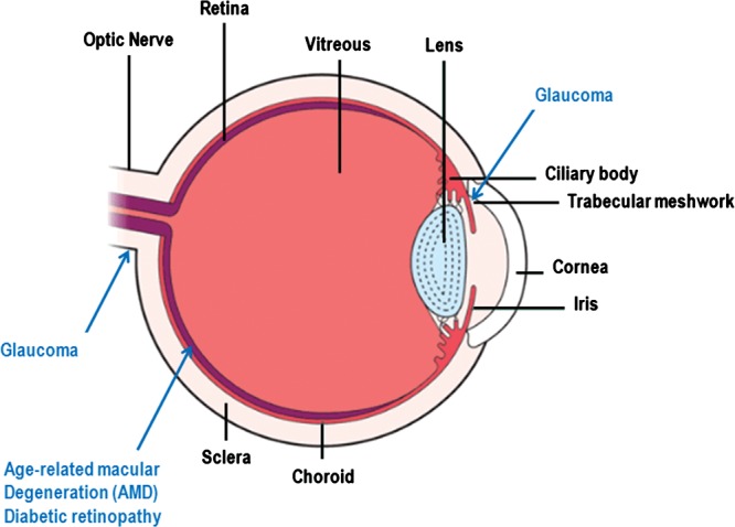
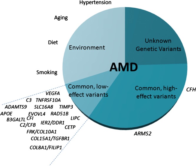
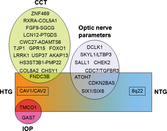
Genome-wide association studies (GWAS) and other genomic technologies have accelerated the discovery of genes and genomic regions contributing to common human ocular disorders with complex inheritance. Age-related macular degeneration (AMD), diabetic retinopathy (DR), glaucoma and myopia account for the majority of visual impairment worldwide. Over 19 genes and/or genomic regions have been associated with AMD. Current investigations are assessing the clinical utility of risk score panels and therapies targeting disease-specific pathways. DR is the leading cause of blindness in the United States and globally is a major cause of vision loss. Genomic investigations have identified molecular pathways associated with DR in animal models which could suggest novel therapeutic targets. Three types of glaucoma, primary-open-angle glaucoma (POAG), angle-closure glaucoma and exfoliation syndrome (XFS) glaucoma, are common age-related conditions. Five genomic regions have been associated with POAG, three with angle-closure glaucoma and one with XFS. Myopia causes substantial ocular morbidity throughout the world. Recent large GWAS have identified >20 associated loci for this condition. In this report, we present a comprehensive overview of the genes and genomic regions contributing to disease susceptibility for these common blinding ocular disorders and discuss the next steps toward translation to effective gene-based screening tests and novel therapies targeting the molecular events contributing to disease.
Figures
References
-
- Eye Diseases Prevalence Research Group. Causes and prevalence of visual impairment among adults in the United States. Arch. Ophthal. 2004;122:477–485. - PubMed
-
- Manolio T.A. Genomewide association studies and assessment of the risk of disease. N. Engl. J. Med. 2010;363:166–176. - PubMed
-
- Friedman D.S., O'Colmain B.J., Muñoz B., Tomany S.C., McCarty C., de Jong P.T., Nemesure B., Mitchell P., Kempen J. Eye Diseases Prevalence Research Group. Prevalence of age-related macular degeneration in the United States. Arch. Ophthalmol. 2004;122:564–572. - PubMed
-
- Clemons T.E., Milton R.C., Klein R., Seddon J.M., Ferris F.L., III Age-Related Eye Disease Study Research Group. Risk factors for the incidence of Advanced Age-Related Macular Degeneration in the Age-Related Eye Disease Study (AREDS) AREDS report no. 19. Ophthalmology. 2005;112:533–539. - PMC - PubMed
Publication types
MeSH terms
Grants and funding
LinkOut - more resources
Full Text Sources
Other Literature Sources
Medical
