

Physical activity is associated with improved aerobic exercise capacity over time in adults with congenital heart disease
- PMID: 23962775
- PMCID: PMC3809326
- DOI: 10.1016/j.ijcard.2013.07.177
Physical activity is associated with improved aerobic exercise capacity over time in adults with congenital heart disease
Abstract
Background: Impaired exercise capacity is common in adults with congenital heart disease (ACHD). This impairment is progressive and is associated with increased morbidity and mortality. We studied the influence of the frequency of at least moderately strenuous physical activity (PhysAct) on changes in exercise capacity of ACHD patients over time.
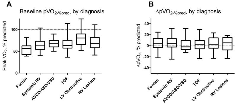
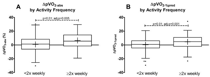
Methods: We studied ACHD patients ≥21 years old who had repeated maximal (RER≥1.09) cardiopulmonary exercise tests within 6 to 24 months. On the basis of data extracted from each patient's clinical records, PhysAct frequency was classified as (1) Low: minimal PhysAct, (2) Occasional: moderate PhysAct <2 times/week, or (3) Frequent: moderate PhysAct ≥2 times/week.
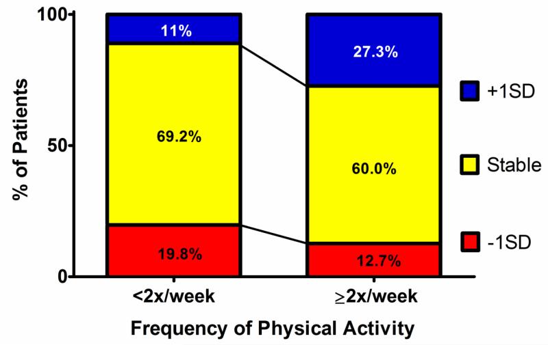
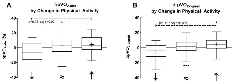
Results: PhysAct frequency could be classified for 146 patients. Those who participated in frequent exercise tended to have improved pVO2 (∆pVO2=+1.63±2.67 ml/kg/min) compared to those who had low or occasional activity frequency (∆pVO2=+0.06±2.13 ml/kg/min, p=0.003) over a median follow-up of 13.2 months. This difference was independent of baseline clinical characteristics, time between tests, medication changes, or weight change. Those who engaged in frequent PhysAct were more likely to have an increase of pVO2 of ≥1SD between tests as compared with sedentary patients (multivariable OR=7.4, 95%CI 1.5-35.7). Aerobic exercise capacity also increased for patients who increased activity frequency from baseline to follow-up; 27.3% of those who increased their frequency of moderately strenuous physical activity had a clinically significant (at least +1SD) increase in pVO2 compared to only 11% of those who maintained or decreased activity frequency.
Conclusions: ACHD patients who engage in frequent physical activity tend to have improved exercise capacity over time.
Keywords: Adults; Congenital heart disease; Exercise capacity and physical activity.
© 2013.
Figures
Similar articles
-
Factors Associated With Increased Exercise in Adults With Congenital Heart Disease.Am J Cardiol. 2019 Sep 15;124(6):947-951. doi: 10.1016/j.amjcard.2019.05.073. Epub 2019 Jun 25. Am J Cardiol. 2019. PMID: 31327487
-
Exercise intolerance in adult congenital heart disease: comparative severity, correlates, and prognostic implication.Circulation. 2005 Aug 9;112(6):828-35. doi: 10.1161/CIRCULATIONAHA.104.529800. Epub 2005 Aug 1. Circulation. 2005. PMID: 16061735
-
Peak oxygen consumption by smartwatches compared with cardiopulmonary exercise test in complex congenital heart disease.Heart. 2024 Feb 12;110(5):353-358. doi: 10.1136/heartjnl-2023-322989. Heart. 2024. PMID: 37827554
-
Examining the Role of Exercise Training in Enhancing Life for Adult Congenital Heart Disease: Systematic Review.Arq Bras Cardiol. 2024 Nov;121(12):e20240294. doi: 10.36660/abc.20240294. Arq Bras Cardiol. 2024. PMID: 39699456 Free PMC article. English, Portuguese.
-
Reference values for exercise limitations among adults with congenital heart disease. Relation to activities of daily life--single centre experience and review of published data.Eur Heart J. 2012 Jun;33(11):1386-96. doi: 10.1093/eurheartj/ehr461. Epub 2011 Dec 23. Eur Heart J. 2012. PMID: 22199119 Review.
Cited by
-
National Trends in Hospitalizations of Adults With Tetralogy of Fallot.Am J Cardiol. 2016 Sep 15;118(6):906-911. doi: 10.1016/j.amjcard.2016.06.034. Epub 2016 Jun 27. Am J Cardiol. 2016. PMID: 27530825 Free PMC article.
-
Emerging hemodynamic signatures of the right heart (Third International Right Heart Failure Summit, part 2).Pulm Circ. 2014 Dec;4(4):705-16. doi: 10.1086/678544. Pulm Circ. 2014. PMID: 25610606 Free PMC article.
-
Growth and Obesity Among Older Single Ventricle Patients Presenting for Fontan Conversion.World J Pediatr Congenit Heart Surg. 2015 Oct;6(4):514-20. doi: 10.1177/2150135115598212. World J Pediatr Congenit Heart Surg. 2015. PMID: 26467864 Free PMC article.
-
Trends and advances in cardiac rehabilitation for congenital heart disease: a bibliometric analysis.J Cardiothorac Surg. 2025 Jul 28;20(1):316. doi: 10.1186/s13019-025-03562-z. J Cardiothorac Surg. 2025. PMID: 40722104 Free PMC article. Review.
-
Overweight predicts poorer exercise capacity in congenital heart disease patients.Int J Cardiol Heart Vasc. 2015 Jul 29;9:28-31. doi: 10.1016/j.ijcha.2015.07.005. eCollection 2015 Dec 7. Int J Cardiol Heart Vasc. 2015. PMID: 28785702 Free PMC article.
References
-
- Marelli AJ, Mackie AS, Ionescu-Ittu R, et al. Congenital heart disease in the general population: changing prevalence and age distribution. Circulation. 2007;115:163–72. - PubMed
-
- Muller J, Christov F, Schreiber C, et al. Exercise capacity, quality of life, and daily activity in the long-term follow-up of patients with univentricular heart and total cavopulmonary connection. Eur Heart J. 2009;30:2915–20. - PubMed
-
- Buys R, Cornelissen V, Van De Bruaene A, et al. Measures of exercise capacity in adults with congenital heart disease. Int J Cardiol. 2011;153:26–30. - PubMed
-
- Fernandes SM, Alexander ME, Graham DA, et al. Exercise testing identifies patients at increased risk for morbidity and mortality following Fontan surgery. Congenit Heart Dis. 2011;6:294–303. - PubMed
Publication types
MeSH terms
Grants and funding
LinkOut - more resources
Full Text Sources
Other Literature Sources
Medical
