. 2014 Mar;134(3):754-763.
doi: 10.1038/jid.2013.356. Epub 2013 Aug 20.

Keratin 9 is required for the structural integrity and terminal differentiation of the palmoplantar epidermis

Affiliations
Free PMC article

Keratin 9 is required for the structural integrity and terminal differentiation of the palmoplantar epidermis

Dun Jack Fu et al. J Invest Dermatol. 2014 Mar.
Free PMC article

Abstract

Keratin 9 (K9) is a type I intermediate filament protein whose expression is confined to the suprabasal layers of the palmoplantar epidermis. Although mutations in the K9 gene are known to cause epidermolytic palmoplantar keratoderma, a rare dominant-negative skin disorder, its functional significance is poorly understood. To gain insight into the physical requirement and importance of K9, we generated K9-deficient (Krt9(-/-)) mice. Here, we report that adult Krt9(-/-)mice develop calluses marked by hyperpigmentation that are exclusively localized to the stress-bearing footpads. Histological, immunohistochemical, and immunoblot analyses of these regions revealed hyperproliferation, impaired terminal differentiation, and abnormal expression of keratins K5, K14, and K2. Furthermore, the absence of K9 induces the stress-activated keratins K6 and K16. Importantly, mice heterozygous for the K9-null allele (Krt9(+/-)) show neither an overt nor histological phenotype, demonstrating that one Krt9 allele is sufficient for the developing normal palmoplantar epidermis. Together, our data demonstrate that complete ablation of K9 is not tolerable in vivo and that K9 is required for terminal differentiation and maintaining the mechanical integrity of palmoplantar epidermis.


Figures
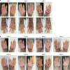
Figure 1
Figure 1
The stress-bearing footpads of fore-paws in keratin 9-deficient (Krt9−/−) mice develop hyperpigmented calluses. The fore- and hind-paws of 8-week old Krt9+/+, Krt9+/−, and Krt9−/− littermates (n=4 per genotype) were photographed weekly over an 8-week period. (a) Initial observation in week 1 revealed hyperpigmented calluses on the major stress-bearing fore-paw footpads of adult Krt9−/− mice (arrows). (b) In Krt9−/− mice, callus size and hyperpigmentation state grew and intensified, respectively, over a ∼3-week period on the major footpads before sloughing off (arrows). Similar, but milder, calluses were also observed on the minor fore-paw footpads of Krt9−/− mice, which also appeared to cycle but at a slower rate (arrowhead).
Figure 2
Figure 2
Localized keratotic phenotypes develop in adult keratin 9-deficient (Krt9−/−) footpads. (a) Hematoxylin and eosin-stained cross-sections of formalin fixed, paraffin-embedded major fore-paw footpads of Krt9+/+, Krt9+/−, and Krt9−/− littermates (n=3 per genotype) revealed acanthosis, hyperkeratosis, and sporadic hypergranulosis in Krt9−/− mice. Granular keratinocytes also failed to compact in these animals and dark pigment granules (circled) were observed in all strata. Scale bar=100 μm. (b) Axiovision imaging software was used to measure the thickness of the footpad and inter-footpad epidermis in hematoxylin and eosin-stained fore-paw cross-sections from Krt9+/+, Krt9+/−, and Krt9−/− animals. Graph depicts the mean thickness of each region calculated from 10 independent measurements taken over the length of a representative section from each genotype (n=3) and the error bars represent SD. ***P⩽0.0001.
Figure 3
Figure 3
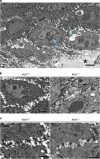
Ultrastructural analysis of keratin 9-deficient (Krt9−/−) fore-paw footpads shows disrupted cytoskeletal integrity and keratin filament assembly. (a) Krt9−/− keratinocytes adjacent to the area of blistering (asterisk) show cytoplasmic disruption with evident cytolysis (green arrow), fewer keratin filaments and some filament, or keratin protein aggregation (blue arrow). Scale bar=1 mm. (b) Compared with Krt9+/+, loss of K9 in leads to relative pallor in Krt9−/− keratinocyte cytoplasm. Scale bar=0.2 mm. (c) Despite loss of K9 (right panel), desmosome cell–cell junctions are similar in size and morphology to Krt9+/+ desmosomes (left panel). Interestingly, fewer keratin filaments are seen in the adjacent cytoplasm in Krt9−/− fore-paw tissues. Both Krt9+/+ and Krt9−/− desmosomes shown here are within the mid-spinous layer. Scale bar=0.1 mm.
Figure 4
Figure 4
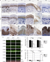
Hyperproliferation in adult keratin 9-deficient (Krt9−/−) footpad epidermis. Immunoperoxidase staining of formalin fixed, paraffin-embedded fore-paw footpads from Krt9+/+, Krt9+/−, and Krt9−/− littermates (n=3 per genotype) with α-Ki-67 (Ki67; MM1-L-CE), α-p63 (p63; SC8431), α-loricrin (LOR; AF62), and α-filaggrin (FLG; gift from Richard Presland, Seattle, WA) antibodies. Note the overall increase in Ki-67-positive basal layer keratinocytes, as well as increased Ki-67 and p63 staining intensities, in the Krt9−/− cross-sections relative to Krt9+/+ and Krt9+/− samples. Although both FLG and LOR localized to the stratum granulosum in all genotypes, LOR staining in Krt9−/− cross-sections was stronger and extended into the stratum corneum. In addition, FLG appeared patchy and disorganized in Krt9−/− samples. Scale bar=50 μm.
Figure 5
Figure 5
Keratin 9-deficient (Krt9−/−)-knockout alters the expression patterns of a subset of palmoplantar keratins. (a) Immunoperoxidase staining of fore-paw footpads from Krt9+/+, Krt9+/−, and Krt9−/− littermates (n=3 per genotype) with α-K5, α-K14, α-K1, α-K10, α-K2, and α-K6. Note increased suprabasal staining of K5 and K14, and increased K6 staining in Krt9−/− cross-sections. Scale bar=50 μm. (b) Immunoblotting of fore-paw footpad whole-tissue lysates Krt9+/+, Krt9+/−, and Krt9−/− littermates (n=3 per genotype) with α-K5, α-K14, α-K1, α-K10, α-K2, α-K6, α-K16, and α-β-actin (loading control; representative image shown) antibodies and differential fluorescence visualization. (c, d) Semiquantitative immunoblotting analyses of α-K5, α-K2, α-K6, and α-K16, relative to β-actin, were used to quantify protein abundance changes following loss of K9. Mean relative abundances (n=3) are shown, and error bars indicate signal intensity SD. *P⩽0.05; **P⩽0.01.

Similar articles

Cited by

References

    1. Candi E, Cipollone R, Rivetti di Val Cervo P, et al. p63 in epithelial development. Cell Mol Life Sci. 2008;65:3126–3133. - PMC - PubMed
    1. Candi E, Schmidt R, Melino G. The cornified envelope: a model of cell death in the skin. Nat Rev Mol Cell Biol. 2005;6:328–340. - PubMed
    1. Chan YM, Ka-Leung Cheng D, Nga-Yin Cheung A, et al. Female urethral adenocarcinoma arising from urethritis glandularis. Gynecol Oncol. 2000;79:511–514. - PubMed
    1. Cook HC.1974Manual of Histological Demonstration TechniquesButterworths, London, 314p.
    1. Coulombe PA, Tong X, Mazzalupo S, et al. Great promises yet to be fulfilled: defining keratin intermediate filament function in vivo. Eur J Cell Biol. 2004;83:735–746. - PubMed

Publication types

LinkOut - more resources