

P2X7 receptor antagonism inhibits p38 mitogen-activated protein kinase activation and ameliorates neuronal apoptosis after subarachnoid hemorrhage in rats
- PMID: 23963136
- PMCID: PMC3841260
- DOI: 10.1097/CCM.0b013e31829a8246
P2X7 receptor antagonism inhibits p38 mitogen-activated protein kinase activation and ameliorates neuronal apoptosis after subarachnoid hemorrhage in rats
Abstract
Objectives: Brilliant blue G, a selective P2X7 receptor antagonist, exhibits neuroprotective properties. This study examined whether brilliant blue G treatment ameliorates early brain injury after experimental subarachnoid hemorrhage, specifically via inhibiting p38 mitogen-activated protein kinase-related proapoptotic pathways.
Design: Controlled in vivo laboratory study.
Setting: Animal research laboratory.
Subjects: One hundred fifty-four adult male Sprague-Dawley rats weighing 280-320 g.
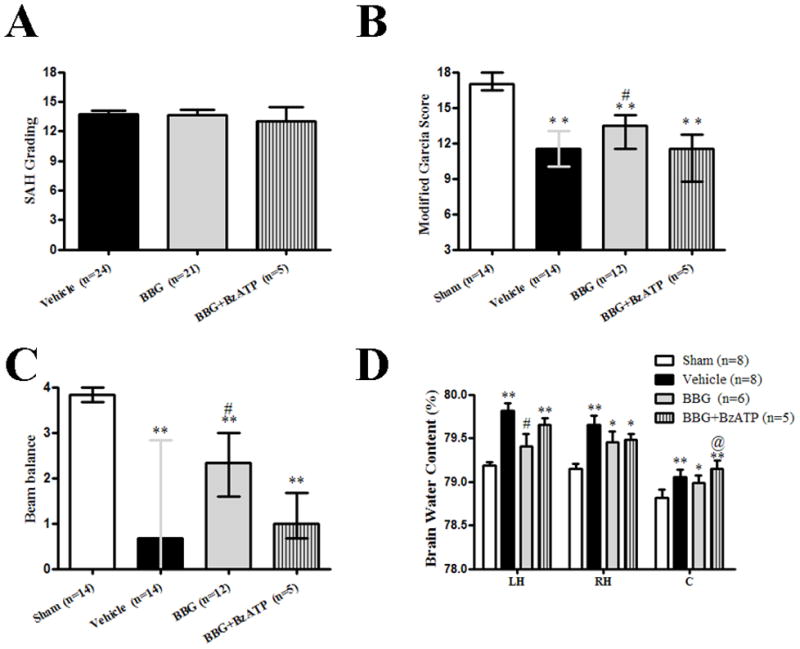
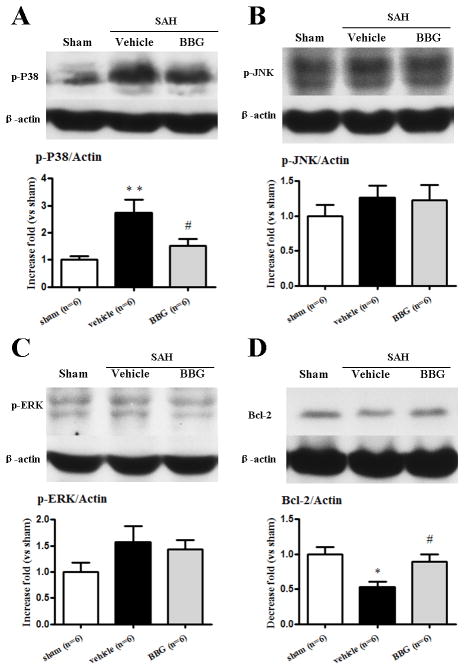
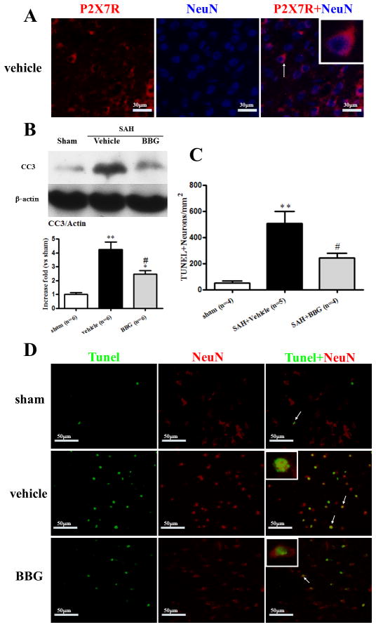
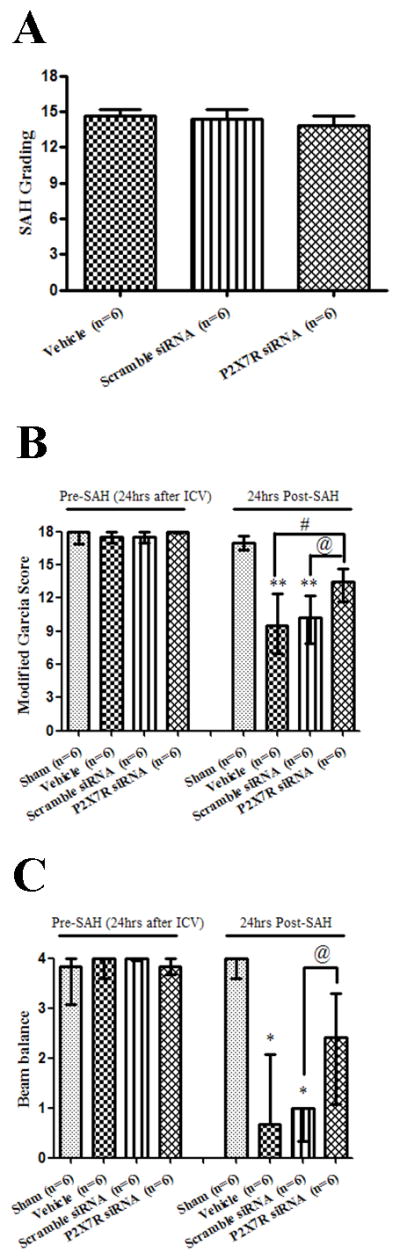
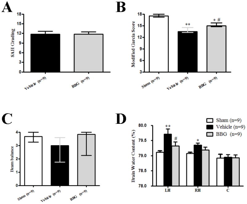
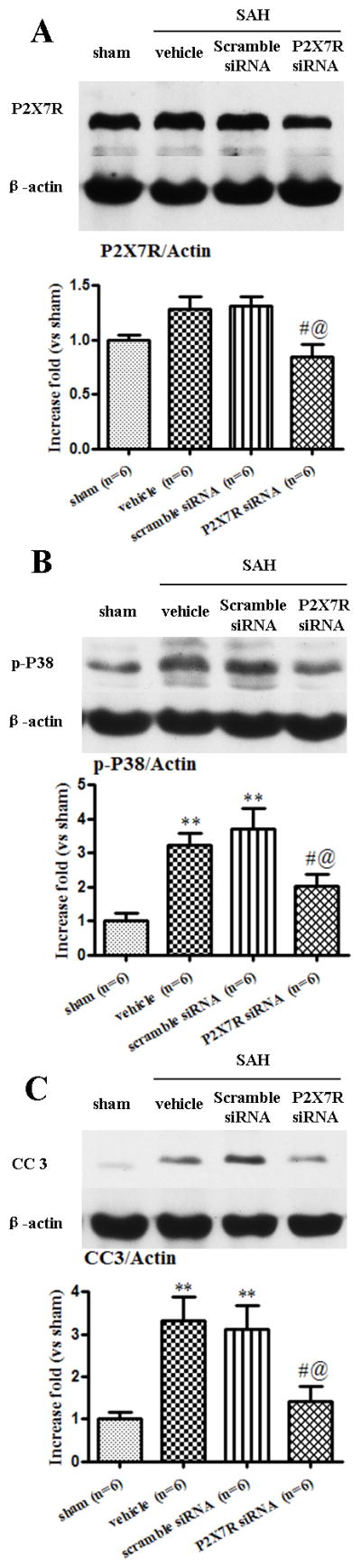
Interventions: Subarachnoid hemorrhage was induced in rats by endovascular perforation. Experiment 1 implemented sham-operated rats (sham) and subarachnoid hemorrhage animals, which received vehicle (subarachnoid hemorrhage + vehicle), brilliant blue G (subarachnoid hemorrhage + brilliant blue G), or brilliant blue G plus 2'(3')-O-(4-Benzoylbenzoyl)adenosine 5'-triphosphate (BzATP) (subarachnoid hemorrhage + brilliant blue G + BzATP). The animals were intraperitoneally treated with brilliant blue G (30 mg/kg) at 30 minutes after subarachnoid hemorrhage. BzATP (50 μg/rat), a P2X7 receptor agonist, was intracerebroventricularly administered. Experiment 2 implemented sham-operated rats (sham) and subarachnoid hemorrhage animals, which received vehicle (subarachnoid hemorrhage + vehicle), scramble small interfering RNA (subarachnoid hemorrhage + scramble small interfering RNA), or P2X7 receptor small interfering RNA (subarachnoid hemorrhage + P2X7 receptor small interfering RNA). Subarachnoid hemorrhage grading, neurobehavioral score, and brain edema were evaluated at 24 and 72 hours after surgery. The expression of phosphorylated p38 mitogen-activated protein kinase, phosphorylated extracellular signal-regulated kinases, phosphorylated c-Jun N-terminal kinases, P2X7 receptor, Bcl-2, and cleaved caspase-3 in the left cerebral hemisphere were determined by Western blot. Neuronal apoptosis was examined by double immunofluorescence staining using P2X7 receptor, terminal deoxynucleotidyl transferase-mediated uridine 5'-triphosphate-biotin nick end-labeling, and neuronal nuclei.
Measurements and main results: Brilliant blue G significantly improved neurobehavioral function and ameliorated brain water content at 24 and 72 hours after subarachnoid hemorrhage. BzATP reversed these treatment effects. Brilliant blue G attenuated neuronal apoptosis in the subcortex, which was associated with decreased expression of phosphorylated p38 mitogen-activated protein kinase and cleaved caspase-3 and an increased expression of Bcl-2 in the left cerebral hemisphere. The beneficial effects of P2X7 receptor small interfering RNA were also mediated by a p38 mitogen-activated protein kinase pathway.
Conclusions: Inhibition of P2X7 receptor by brilliant blue G or P2X7 receptor small interfering RNA can prevent early brain injury via p38 mitogen-activated protein kinase after subarachnoid hemorrhage.
Conflict of interest statement
Conflicts of Interest: None.
Figures
Comment in
-
P2X7 receptor and apoptosis.Crit Care Med. 2014 Dec;42(12):e804. doi: 10.1097/CCM.0000000000000621. Crit Care Med. 2014. PMID: 25402310 No abstract available.
-
The authors reply.Crit Care Med. 2014 Dec;42(12):e804-5. doi: 10.1097/CCM.0000000000000652. Crit Care Med. 2014. PMID: 25402311 Free PMC article. No abstract available.
References
-
- Venti M. Subarachnoid and intraventricular hemorrhage. Front Neurol Neurosci. 2012;30:149–153. - PubMed
-
- Hasegawa Y, Suzuki H, Sozen T, et al. Apoptotic mechanisms for neuronal cells in early brain injury after subarachnoid hemorrhage. Acta Neurochir Suppl. 2011;110:43–48. - PubMed
-
- Skaper SD, Debetto P, Giusti P. The P2X7 purinergic receptor: from physiology to neurological disorders. FASEB J. 2010;24:337–345. - PubMed
-
- Takenouchi T, Sekiyama K, Sekigawa A, et al. P2X7 receptor signaling pathway as a therapeutic target for neurodegenerative diseases. Arch Immunol Ther Exp (Warsz) 2010;58:91–96. - PubMed
Publication types
MeSH terms
Substances
Grants and funding
LinkOut - more resources
Full Text Sources
Other Literature Sources
Research Materials
Miscellaneous
