. 2013:3:2459.
doi: 10.1038/srep02459.

Silibinin suppresses EMT-driven erlotinib resistance by reversing the high miR-21/low miR-200c signature in vivo

Affiliations

Silibinin suppresses EMT-driven erlotinib resistance by reversing the high miR-21/low miR-200c signature in vivo

Sílvia Cufí et al. Sci Rep. 2013.

Abstract

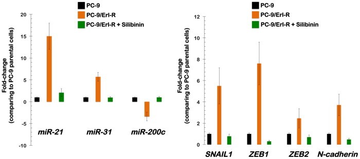
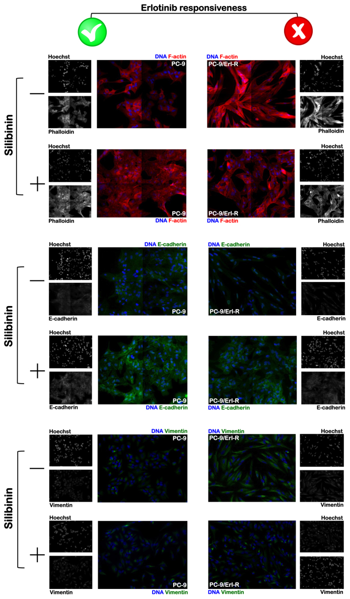
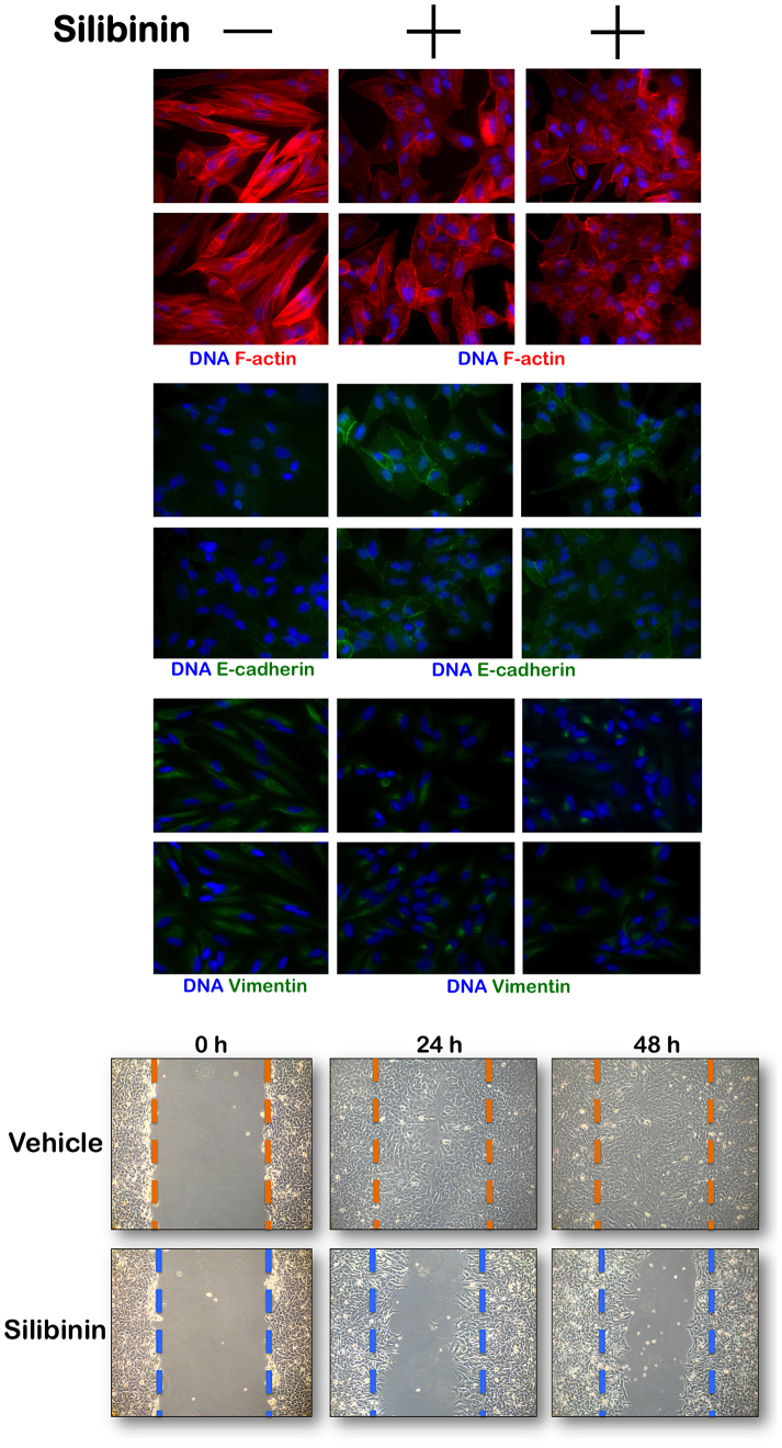
The flavolignan silibinin was studied for its ability to restore drug sensitivity to EGFR-mutant NSCLC xenografts with epithelial-to-mesenchymal transition (EMT)-driven resistance to erlotinib. As a single agent, silibinin significantly decreased the tumor volumes of erlotinib-refractory NSCLC xenografts by approximately 50%. Furthermore, the complete abrogation of tumor growth was observed with the co-treatment of erlotinib and silibinin. Silibinin fully reversed the EMT-related high miR-21/low miR-200c microRNA signature and repressed the mesenchymal markers SNAIL, ZEB, and N-cadherin observed in erlotinib-refractory tumors. Silibinin was sufficient to fully activate a reciprocal mesenchymal-to-epithelial transition (MET) in erlotinib-refractory cells and prevent the highly migratogenic phenotype of erlotinib-resistant NSCLC cells. Given that the various mechanisms of resistance to erlotinib result from EMT, regardless of the EGFR mutation status, a water-soluble, silibinin-rich milk thistle extract might be a suitable candidate therapy for upcoming clinical trials aimed at preventing or reversing NSCLC progression following erlotinib treatment.


Conflict of interest statement

AstraZeneca (Spain) provided partial financial support for the present study via an educational grant to Dr. Javier A. Menendez and Dr. Joaquim Bosch-Barrera.

Figures

Figure 1
Figure 1. HPLC chromatogram of milk thistle extract with 30% (w/w) of water-soluble silibinin meglumine at 288 nm.
Compounds are identified by number as follows: silychristin A (1), silydianin (2), silychristin B (3), silybin A (4), silybin B (5), isosilybin A (6) and isosilybin B (7). Chemical structures of the compounds are also shown.
Figure 2
Figure 2. Oral treatment of erlotinib-refractory, EGFR-mutant NSCLC xenograft-bearing animals with silibinin: Impact on the efficacy of the EGFR TKI erlotinib in vivo.
(A). Shown are the mean tumor volumes (±SD) in PC-9/Erl-R xenograft-bearing nude mice following oral gavage with erlotinib, silibinin or erlotinib plus silibinin for five weeks. Data represent means ± SD. *P < 0.05 (Student's t-test), **P < 0.005 (Student's t-test), and n.s. non-statistically significant results (Student's t-test), versus erlotinib-treated mice. (B). Antitumor activity was calculated for individual tumors as the percentage of tumor growth inhibition, according to the following formula: 100 − [(Vx/Vc) × 100], where Vx is the tumor volume for treated mice and Vc is the tumor volume in the control group at a given x time.
Figure 3
Figure 3. Oral treatment with silibinin reverses the EMT-related high miR-21/low miR200c microRNA signature and downregulates mesenchymal markers in erlotinib-refractory EGFR-mutant NSCLC xenograft-bearing animals.
Figure shows the difference (fold-change mean values ± SD versus untreated PC-9 parental cells; n = 4) in the three differentially expressed miRNAs (i.e., miR-21, miR-31, and miR-200c) between erlotinib-resistant PC-9/Erl-R tumor samples and erlotinib-responsive PC-9 tumor samples following oral treatment with silibinin for five weeks (left panel). Figure also shows the effects of oral treatment with silibinin in the expression of the mesenchymal markers SNAIL1, ZEB1, ZEB2, and N-cadherin (fold-change mean values ± SD versus untreated PC-9 parental cells; n = 4), which were found significantly overrepresented in erlotinib-refractory EGFR-mutant NSCLC xenografts (right panel).
Figure 4
Figure 4. Silibinin enhances the epithelial phenotype in PC-9 parental cells and reverses the mesenchymal phenotype in PC-9/Erl-R cells (I).
PC-9 parental cells and erlotinib-resistant PC-9/Erl-R cells were cultured for 3 days in the absence or presence of 100 μg/mL of silibinin-meglumine. Top panels. After fixation and permeabilization, the subcellular distribution of F-actin, the epithelial marker E-cadherin, and of the mesenchymal-specific marker vimentin was assessed after staining with anti-F-actin, anti-E-cadherin, and anti-vimentin antibodies and Hoechst 33258 for nuclear counterstaining, as specified. Images show representative portions of the untreated- and silibinin meglumine-treated cells in montages of 4 × 4, which were captured in different channels for F-actin (red), E-cadherin (green), vimentin (green), and Hoechst 33258 (blue) with a 20× objective. Images were merged on a BD Pathway 855 Bioimager System with BD Attovision software.
Figure 5
Figure 5. Silibinin promotes mesenchymal to epithelial conversion of erlotinib-resistant PC-9/Erl-R cells.
Top panels. Erlotinib-resistant PC-9/Erl-R cells were cultured for 3 days in the absence or presence of 100 μg/mL of silibinin. Top panels. After fixation and permeabilization, the subcellular distribution of F-actin, the epithelial marker E-cadherin, and of the mesenchymal-specific marker vimentin was assessed after staining with anti-F-actin, anti-E-cadherin, and anti-vimentin antibodies and Hoechst 33258 for nuclear counterstaining, as specified. Images show representative portions of the untreated- and silibinin meglumine-treated cells, which were captured in different channels for F-actin (red), E-cadherin (green), vimentin (green), and Hoechst 33258 (blue) with a 20× objective. Images were merged on a BD Pathway 855 Bioimager System with BD Attovision software. Bottom panels. Figure shows representative microphotographs of wound healing assays 0, 24, and 48 h after incisions of confluent PC-9/ErL-R cells cultured in the absence or presence of silibinin (100 μg/mL).
Figure 6
Figure 6. Water-soluble silibinin: A new strategy for targeted in vivo control of molecules that regulate the acquisition of an EGFR TKI-refractory mesenchymal-like phenotype in EGFR mutant-NSCLC cells.

References

    1. Rosell R. et al. Screening for epidermal growth factor receptor mutations in lung cancer. N Engl J Med. 361, 958–67 (2009). - PubMed
    1. Rosell R. et al. Predictive biomarkers in the management of EGFR mutant lung cancer. Ann N Y Acad Sci 1210, 45–52 (2010). - PubMed
    1. Rosell R. et al. Non-small-cell lung cancer harbouring mutations in the EGFR kinase domain. Clin Transl Oncol 12, 75–80 (2010). - PubMed
    1. Rosell R. et al. Erlotinib versus standard chemotherapy as first-line treatment for European patients with advanced EGFR mutation-positive non-small-cell lung cancer (EURTAC): a multicentre, open-label, randomised phase 3 trial. Lancet Oncol 13, 239–46 (2012). - PubMed
    1. Santarpia M., De Pas T. M., Altavilla G., Spaggiari L. & Rosell R. Moving towards molecular-guided treatments: erlotinib and clinical outcomes in non-small-cell lung cancer patients. Future Oncol 9, 327–45 (2013). - PubMed

Publication types

MeSH terms