

Deletions involving genes WHSC1 and LETM1 may be necessary, but are not sufficient to cause Wolf-Hirschhorn Syndrome
- PMID: 23963300
- PMCID: PMC3953918
- DOI: 10.1038/ejhg.2013.192
Deletions involving genes WHSC1 and LETM1 may be necessary, but are not sufficient to cause Wolf-Hirschhorn Syndrome
Abstract
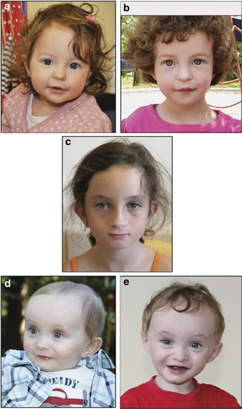
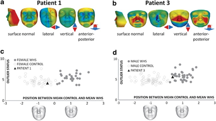
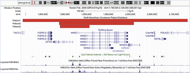
Wolf-Hirschhorn syndrome (WHS) is a complex genetic disorder caused by the loss of genomic material from the short arm of chromosome 4. Genotype-phenotype correlation studies indicated that the loss of genes within 4p16.3 is necessary for expression of the core features of the phenotype. Within this region, haploinsufficiency of the genes WHSC1 and LETM1 is thought to be a major contributor to the pathogenesis of WHS. We present clinical findings for three patients with relatively small (<400 kb) de novo interstitial deletions that overlap WHSC1 and LETM1. 3D facial analysis was performed for two of these patients. Based on our findings, we propose that hemizygosity of WHSC1 and LETM1 is associated with a clinical phenotype characterized by growth deficiency, feeding difficulties, and motor and speech delays. The deletion of additional genes nearby WHSC1 and LETM1 does not result in a marked increase in the severity of clinical features, arguing against their haploinsufficiency. The absence of seizures and typical WHS craniofacial findings in our cohort suggest that deletion of distinct or additional 4p16.3 genes is necessary for expression of these features. Altogether, these results show that although loss-of-function for WHSC1 and/or LETM1 contributes to some of the features of WHS, deletion of additional genes is required for the full expression of the phenotype, providing further support that WHS is a contiguous gene deletion disorder.
Figures
References
-
- Maas NM, Van Buggenhout G, Hannes F, et al. Genotype-phenotype correlation in 21 patients with Wolf-Hirschhorn syndrome using high resolution array comparative genome hybridisation (CGH) J Med Genet. 2008;45:71–80. - PubMed
-
- Battaglia A, Carey JC, South ST, Wright TJ.Wolf-Hirschhorn syndromein: Pagon RA, Bird TD, Dolan CR, Stephens K, Adam MP, (eds): GeneReviews Seattle, WA, USA: University of Washington, Seattle; 1993 - PubMed
-
- Battaglia A, Carey JC, Wright TJ. Wolf-Hirschhorn (4p-) syndrome. Adv Pediatr. 2001;48:75–113. - PubMed
-
- Battaglia A, Filippi T, Carey JC. Update on the clinical features and natural history of Wolf-Hirschhorn (4p-) syndrome: experience with 87 patients and recommendations for routine health supervision. Am J Med Genet C Sem Med Genet. 2008;148C:246–251. - PubMed
-
- South ST, Whitby H, Battaglia A, Carey JC, Brothman AR. Comprehensive analysis of Wolf-Hirschhorn syndrome using array CGH indicates a high prevalence of translocations. Eur J Hum Genet. 2008;16:45–52. - PubMed
Publication types
MeSH terms
Substances
Grants and funding
LinkOut - more resources
Full Text Sources
Other Literature Sources
