. 2013 Oct 4;288(40):28620-9.
doi: 10.1074/jbc.M113.452169. Epub 2013 Aug 20.

Transcriptional induction of ADAMTS5 protein by nuclear factor-κB (NF-κB) family member RelA/p65 in chondrocytes during osteoarthritis development

Affiliations

Transcriptional induction of ADAMTS5 protein by nuclear factor-κB (NF-κB) family member RelA/p65 in chondrocytes during osteoarthritis development

Hiroshi Kobayashi et al. J Biol Chem. .

Abstract

Here we sought to identify transcription factors that induce ADAMTS5, a crucial proteinase for osteoarthritis development. Exhaustive comparison of the genomic sequences of human, macaque, and mouse ADAMTS5 genes revealed that the proximal 1.4 kb of the 5'-end-flanking regions containing several consensus motifs was highly conserved. Among putative transcription factors for these motifs, NF-κB family member RelA/p65 most strongly stimulated the promoter activity. In the ADAMTS5 gene, there were three NF-κB binding motifs, in which deletion, mutagenesis, and tandem repeat analyses of the luciferase assay identified the core responsive elements of RelA/p65 to be -896/-887 and -424/-415 bp with specific bindings. The endogenous Adamts5 expression in ATDC5 cells was increased by RelA/p65 overexpression and decreased by knockdown through its siRNA. The expression was also inhibited by the Rela deletion through Cre transfection in primary articular chondrocytes from Rela(fl/fl) mice. In the ex vivo culture of femoral head cartilage from mesenchymal cell-specific Rela knock-out (Prx1-Cre;Rela(fl/fl)) mice, aggrecanolysis was significantly lower than that in the Rela(fl/fl) cartilage. Finally, in the experimental mouse osteoarthritis model, ADAMTS5 and RelA were co-localized in chondrocytes of degraded articular cartilage. We conclude that RelA/p65 is a potent transcriptional activator of ADAMTS5 in chondrocytes during osteoarthritis development.

Keywords: ADAM ADAMTS; ADAMTS5; Articular Cartilage; Cartilage; Cartilage Biology; Chondrocytes; NF-kappa B (NF-KB); Osteoarthritis; RelA/p65; Transcription.


Figures
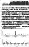
FIGURE 1.
FIGURE 1.
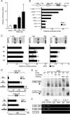
Identification of transcription factors of the ADAMTS5 promoter. A, the sequence similarity of the proximal promoter (2 kb) sequences among the mouse, macaque, and human ADAMTS5 genes analyzed by BLASTN search and Vector-NTI software (Invitrogen). B, conserved nucleotides in the two or three species are denoted by white letters shaded in gray or black, respectively. An arrow shows the TSS of the human gene. Putative binding motifs of transcription factors identified in highly conserved regions are indicated below. C, luciferase assay for the human ADAMTS5 promoter activity by the putative transcription factors whose binding motifs were identified above. ATDC5 cells were co-transfected with a luciferase reporter construct containing the 5′-end-flanking region (−1,400/+1) of the human ADAMTS5 gene and the effecter vectors or the control empty vector (EV). *, p < 0.05, **, p < 0.01 versus EV. D, luciferase assay for the human ADAMTS5 promoter activity by the co-transfection of putative binding partners with RelA/p65 in the reporter construct containing ATDC5 cells as above. For C and D, data are shown as means (bars) ± S.E. (error bars) of relative luciferase activity (the ratio of the firefly activities to the Renilla activities) for three wells/group.
FIGURE 2.
FIGURE 2.
Identification of the core region responsive to RelA/p65 in the ADAMTS5 proximal promoter. A, dose response of the ADAMTS5 promoter activity with different amounts (25, 50, and 75 μg) of the RelA/p65 expression vector in the ATDC5 cells co-transfected with a luciferase reporter construct containing the 5′-end-flanking region (−1,400/+1) of the human ADAMTS5 gene. *, p < 0.05 versus EV. B, deletion analysis of the luciferase assay in ATDC5 cells co-transfected with a luciferase reporter construct containing a series of deletion fragments of the human ADAMTS5 promoter including three NF-κB motifs (−1,196/−1,187, −896/−887, and −424/−415) and human RelA/p65 or the control EV. *, p < 0.05 versus −1,022/+1 fragment with RelA. #, p < 0.05 versus −506/+1 fragment with RelA. C, mutagenesis analysis of the luciferase assay in ATDC5 cells co-transfected with a luciferase reporter construct containing wild-type (WT) or a single and double mutations in the NF-κB motifs (M1, M2, and M3 in one motif), and RelA or EV. *, p < 0.05, **, p < 0.01 versus WT+RelA. D, tandem repeat analysis of the luciferase assay in ATDC5 cells co-transfected with a luciferase reporter construct ligated to the −896/−887 motif (upper) or to the −424/−415 motif (lower), and RelA or EV. *, p < 0.05, **, p < 0.01 versus ×1 fragment with RelA. For A–D, data are shown as means (bars) ± S.E. (error bars) of relative luciferase activity for three wells/group. E, EMSA for binding of nuclear extracts (NE) of COS-7 cells transfected with RelA or the control empty vector (−) and the WT or the mutated (M1) probe above of the −896/−887 (top) and the −424/−415 (bottom) oligonucleotide probes. The arrowheads indicate the protein-probe binding. Cold competition with 50-fold excess of unlabeled WT or M1 probe is presented. A supershift of the binding by an antibody to RelA or nonimmune IgG is also shown. F, ChIP assay for specific binding of RelA to the NF-κB motif using cell lysates of human chondrogenic OUMS-27 cells with or without IL-1β treatment. Primer sets that amplify the area including the NF-κB motif (−896/−887 or −424/−415) or that do not included the motif (+17/+213) were employed before (input) and after immunoprecipitation with an antibody to RelA or the control nonimmune IgG.
FIGURE 3.
FIGURE 3.
Effects of gain and loss of functions of RelA/p65 on ADAMTS5 expression. A, mRNA levels of Adamts5 and RelA (graphs) and protein levels of RelA (Western blotting) in cultured ATDC5 cells with adenoviral overexpression of RelA/p65 (Ax-RelA) or the control GFP (Ax-GFP). **, p < 0.01 versus Ax-GFP. B, mRNA levels of Adamts5 and Rela in ATDC5 cells transfected with siRNA of RelA (siRela) or the control GFP (siGFP), which were cultured in the presence and absence of IL-1β for 24 h. **, p < 0.01 versus siGFP+control. ##, p < 0.01 versus siGFP+IL-1β. C, mRNA levels of Adamts5 and Rela in primary articular chondrocytes derived from Relafl/fl mice with adenoviral transfection of several concentrations (10, 50, and 100 multiplicity of infection) of Cre of the control GFP. *, p < 0.05, **, p < 0.01 versus Ax-GFP. In all experiments, data are shown as means (bars) ± S.E. (error bars) of three cultures/group.
FIGURE 4.
FIGURE 4.
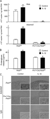
Effects of loss of functions of RelA on aggrecanolysis. A, Rela and Adamts5 mRNA levels in primary chondrocytes of articular cartilage derived from Prx1-Cre;Relafl/fl mice and the control Relafl/fl littermates in the presence and absence of IL-1β for 24 h. *, p < 0.05, **, p < 0.01 versus Relafl/fl+control. #, p < 0.05, ##, p < 0.01 versus Relafl/fl+IL-1β. B, the concentration of total glycosaminoglycan released into the medium in the ex vivo cultures of femoral head cartilage derived from the two genotype mice above with and without IL-1β for 72 h. *, p < 0.05 versus Relafl/fl+control. #, p < 0.05 versus Relafl/fl+IL-1β. For A and B, data are shown as means (bars) ± S.E. (error bars) of three cultures/group using two mice/genotype. C, representative images of immunohistochemical staining by an antibody to ADAMTS5 or the control nonimmune IgG in the cultured femoral head cartilages above (images with high magnification are inserted). Positive stainings are brown by diaminobenzidine with methyl green counterstaining. Scale bars, 20 μm. Immunohistochemistry was performed at least three times using two mice/group/genotype and obtained reproducible results.
FIGURE 5.
FIGURE 5.
Immunohistochemistry using antibodies to RelA and ADAMTS5 and the merged staining, as well as the control nonimmune IgG in various regions of the joint cartilage (the superficial layer with high degeneration, the superficial layer with low degeneration, and the deep layer) of the knee joint cartilage of the experimental mouse OA model. The model was created by surgical induction of instability in the knee joints of 8-week-old mice, and the staining was performed 8 weeks after surgery. The boxed areas in the Safranin-O staining indicate regions shown in the enlarged images of lower panels. Scale bars, 100 μm. These analyses were performed at least three times using three mice and obtained reproducible results.

References

    1. Verma P., Dalal K. (2011) ADAMTS-4 and ADAMTS-5: key enzymes in osteoarthritis. J. Cell. Biochem. 112, 3507–3514 - PubMed
    1. Brophy R. H., Rai M. F., Zhang Z., Torgomyan A., Sandell L. J. (2012) Molecular analysis of age and sex-related gene expression in meniscal tears with and without a concomitant anterior cruciate ligament tear. J. Bone Joint Surg. Am. 94, 385–393 - PMC - PubMed
    1. Glasson S. S., Askew R., Sheppard B., Carito B., Blanchet T., Ma H. L., Flannery C. R., Peluso D., Kanki K., Yang Z., Majumdar M. K., Morris E. A. (2005) Deletion of active ADAMTS5 prevents cartilage degradation in a murine model of osteoarthritis. Nature 434, 644–648 - PubMed
    1. Stanton H., Rogerson F. M., East C. J., Golub S. B., Lawlor K. E., Meeker C. T., Little C. B., Last K., Farmer P. J., Campbell I. K., Fourie A. M., Fosang A. J. (2005) ADAMTS5 is the major aggrecanase in mouse cartilage in vivo and in vitro. Nature 434, 648–652 - PubMed
    1. Glasson S. S., Askew R., Sheppard B., Carito B. A., Blanchet T., Ma H. L., Flannery C. R., Kanki K., Wang E., Peluso D., Yang Z., Majumdar M. K., Morris E. A. (2004) Characterization of and osteoarthritis susceptibility in ADAMTS-4-knockout mice. Arthritis Rheum. 50, 2547–2558 - PubMed

Publication types

MeSH terms