. 2013 Oct 4;288(40):29170-81.
doi: 10.1074/jbc.M113.456947. Epub 2013 Aug 20.

Homotypic versican G1 domain interactions enhance hyaluronan incorporation into fibrillin microfibrils

Affiliations

Homotypic versican G1 domain interactions enhance hyaluronan incorporation into fibrillin microfibrils

Yusuke Murasawa et al. J Biol Chem. .

Abstract

Versican G1 domain-containing fragments (VG1Fs) have been identified in extracts from the dermis in which hyaluronan (HA)-versican-fibrillin complexes are found. However, the molecular assembly of VG1Fs in the HA-versican-microfibril macrocomplex has not yet been elucidated. Here, we clarify the role of VG1Fs in the extracellular macrocomplex, specifically in mediating the recruitment of HA to microfibrils. Sequential extraction studies suggested that the VG1Fs were not associated with dermal elements through HA binding properties alone. Overlay analyses of dermal tissue sections using the recombinant versican G1 domain, rVN, showed that rVN deposited onto the elastic fiber network. In solid-phase binding assays, rVN bound to isolated nondegraded microfibrils. rVN specifically bound to authentic versican core protein produced by dermal fibroblasts. Furthermore, rVN bound to VG1Fs extracted from the dermis and to nondenatured versican but not to fibrillin-1. Homotypic binding of rVN was also seen. Consistent with these binding properties, macroaggregates containing VG1Fs were detected in high molecular weight fractions of sieved dermal extracts and visualized by electron microscopy, which revealed localization to microfibrils at the microscopic level. Importantly, exogenous rVN enhanced HA recruitment both to isolated microfibrils and to microfibrils in tissue sections in a dose-dependent manner. From these data, we propose that cleaved VG1Fs can be recaptured by microfibrils through VG1F homotypical interactions to enhance HA recruitment to microfibrils.

Keywords: ADAM ADAMTS; Dermis; Extracellular Matrix; Fibrillin; Hyaluronate; Proteoglycan; Versican.


Figures
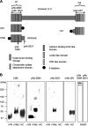
FIGURE 1.
FIGURE 1.
Schematic and characterization of recombinant versican polypeptides and antibodies used in this study. A, schematic of human versican (V1) is shown. Recombinant versican polypeptides, rVN, rVNβ, and rVC are designed as indicated. The recombinant polypeptide rVNβ consists of the G1 domain and the N-terminal portion of the chondroitin sulfate β domain, which ends with the amino acid sequence DPEAAE441 and represents the cleavage site utilized by ADAMTS-1, ADAMTS-4, and ADAMTS-5. Recognition sites for pAb 6084, pAb 7080, pAb 8531, mAb 12C5, and mAb 2B1 are indicated. B, the purified recombinant polypeptides rVN, rVNβ, rVC, and conditioned medium from normal dermal fibroblasts treated with the chondroitinase ABC were subjected to SDS-PAGE under nonreducing conditions and detected by Coomassie Brilliant Blue (CBB) and immunoblotting. Antibodies used for detection are indicated at the top. CRP, complement regulatory protein.
FIGURE 2.
FIGURE 2.
Characterization of VG1Fs from dermal tissue. A, Western blot analyses using anti-versican antibodies and blot overlay assays by bHA. The 6 m guanidine hydrochloride dermal extract was treated with chondroitinase ABC and resolved by SDS-PAGE under nonreducing conditions. The blots were incubated with bHA or specific antibodies as indicated. The blot with pAb 8531 (anti-DPEAAE) showed multiple molecules (arrows) containing the bands larger than 150 kDa (asterisk) B and C, Western blot analyses with pAb 8531. The procedures for extraction from the dermis are indicated and are described in detail under “Experimental Procedures.” B, dermal tissues were sequentially extracted with 6 m guanidine hydrochloride, PBS, and Streptomyces HAase treatment. C, dermal tissues were sequentially extracted with PBS, Streptomyces HAase, and 6 m guanidine hydrochloride. The extracts were precipitated, treated with chondroitinase ABC, resolved on 7.5% acrylamide gels under nonreducing conditions, and blotted.
FIGURE 3.
FIGURE 3.
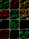
Tissue overlay assays using recombinant VG1Fs. Dermal sections were overlaid with recombinant versican G1 polypeptides. A, bound rVN was detected by FITC-conjugated anti-C-His antibodies (green). B, the endogenous G3 domain was detected by mAb 2B1 (red). C, colocalization of the exogenous rVN and endogenous G3 domain is shown in the merged image (yellow). A–C, scale bars = 20 μm. D, bound rVN was detected by Alexa Fluor 555-conjugated anti-C-His antibodies (red). E, endogenous fibrillin-1 was detected by pAb 9543 (green). F, colocalization of exogenous rVN and endogenous fibrillin-1 is shown in the merged image (yellow). G, the merged image of a control section (treated similarly but without rVN and pAb 9543) was negative. D–G, scale bars = 10 μm. H, In another field of the section, exogenous rVN was detected (green), whereas in I, control sections (treated similarly but without rVN) were negative. J, bound rVN (red) was also detected by Alexa Fluor 568-conjugated antibodies. K, the section overlaid with reduced and alkylated rVN was negative. L, exogenously added rVNβ was similarly detected on tissue sections (green). H–L, scale bars = 20 μm.
FIGURE 4.
FIGURE 4.
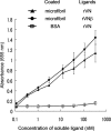
Binding of VG1Fs to extracted microfibrils. Isolated microfibrils extracted from human fetal membranes or bovine serum albumin (□) were immobilized. Coated microfibrils were prepared using 6 m guanidine extraction, molecular sieve chromatography, and ultracentrifugation containing cesium chloride. rVN (●) and rVNβ (▴) were used as soluble ligands. Each point represents the mean ± S.D. obtained from three independent experiments.
FIGURE 5.
FIGURE 5.
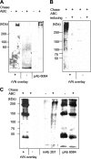
The presence of ligands for rVN in secreted molecules from dermal fibroblasts and in dermal extracts. To identify ligands for rVN, blot overlay assays were performed. Conditioned medium from NHDFs (A and B) and dermal extracts, prepared using 6 m guanidine hydrochloride (C) with (+) or without (-) chondroitinase ABC treatment, were resolved and blotted. A, biotin-conjugated rVN was used as the soluble ligand. bound rVN was detected by HRP-conjugated avidin. Western blot analysis using pAb 6084 (anti-G1) is shown, indicating the migration of the versican core protein. B, anti-His antibodies were used for detection of bound rVN. The samples were run under nonreducing (-) or reducing (+) conditions. C, the dermal extract was the immobilized ligand. Biotin-conjugated rVN was used as the soluble ligand. Western blot analyses using pAb 6084 and mAb 2B1 (Fig. 1) showed species containing N-terminal and C-terminal domains of versican present in dermal extracts.
FIGURE 6.
FIGURE 6.
Interactions of recombinant globular domains of versican. Solid-phase binding assays were performed using rVN as a soluble ligand (A–C). A, native versican purified from NHDF (●) or BSA (□) was immobilized. Reduced and alkylated rVN (■) was also used as a negative control (indicated as rVN(r)). B, native versican, fibrillin-1 (N) (rF11, fibrillin-1 amino-terminal half), or fibrillin-1 (C) (rF6, fibrillin-1 carboxyl-terminal half) was used to coat wells. Soluble rVN bound to native versican (●). In contrast, rVN did not bind to fibrillin-1 (▵) or fibrillin-1 (C) (×). C, biotin-conjugated rVN bound to wells coated with rVN (♦) and to rVC (recombinant G3 domain, ▴) but not to BSA (□). Binding of soluble biotin-conjugated rVN to immobilized BSA was used as a negative control. Each point in A–C is the mean ± S.D. obtained from three independent experiments. D and E, BIAcore sensorgrams of rVN interactions. Experiments were performed with a series of titrated analytes (rVN) in solution with the indicated concentrations, flowed over a CM5 sensor chip immobilized with 1200 RU of rVN (D) or rF6 (E). The analytes were injected at the point indicated by ▵, and the dissociation phase began at the point indicated by ▴. RU, resonance unit.
FIGURE 7.
FIGURE 7.
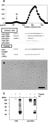
Identification of VG1F-containing aggregates in dermal extracts. A, dermal tissues were extracted with a 6 m guanidine solution. The extract was concentrated using a 50-kDa molecular mass cutoff membrane and sieved on Sepharose CL-2B columns under dissociative conditions. Protein concentrations (micrograms/milliliters), dot blot analyses, and overlay assays using rVN and bHA as soluble ligands are shown. Antibodies used for dot blot analyses are indicated. The void volume fractions were positive with pAb 8531 (neoepitope, DPEAAE) and pAb 7080 (anti-G1 domain) but not with anti-G3 domain antibodies (mAb 2B1) or anti fibrillin-1 antibodies (pAb 9543). Blot overlay analyses using rVN, rVN(r), biotin-conjugated HABP (bHABP), and bHA are also shown. The void fractions bound to rVN and bHA but were negative for rVN(r) and HABP. B, rotary shadowed electron microscopy of VG1F-containing aggregates is shown. The pAb 6084 positive-void volume fractions from sieved chromatography (A) were further isolated by ultracentrifugation containing cesium chloride. Scale bar = 100 nm. C, analysis of the fractions shown in B. Fractions were treated with trypsin alone or trypsin and V8 protease. The samples were resolved by SDS-PAGE, analyzed by staining with Coomassie Brilliant Blue (CBB), and immunoblotting with pAb 6084 (anti-G1).
FIGURE 8.
FIGURE 8.
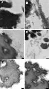
Immunoelectron microscopic localization of the versican G1 domain in the dermal connective tissue. In human adult dermis, pAb 6084 was used to label the versican G1 domain with gold (A and B). The dermis was also incubated with the neoepitope antibody pAb 8531 (C and D) and the anti-versican G3 antibody mAb 2B1 (E). Incubation with nonimmune rabbit serum was performed as a control (F). Scale bars = 100 nm.
FIGURE 9.
FIGURE 9.
Exogenous versican G1 fragments enhance HA deposition into microfibrils. Solid-phase binding assays were performed using bHA as a soluble ligand and immobilized microfibrils bound with rVN (A and B). A, before incubation with hyaluronan, the immobilized microfibrils were incubated with rVN at a constant concentration of 9 μg/ml (●). Binding of bHA was enhanced by the preincubation of exogenous rVN compared with microfibrils without preincubated rVN (■). bHA did not bind to the coated BSA preincubated with rVN (▴). B, solid-phase binding assay using bHA as a soluble ligand at a constant concentration (5 μg/ml). Immobilized microfibrils or BSA were preincubated with increasing concentrations of rVN. C and D, tissue overlay assay using bHA (green) as a soluble ligand on dermal tissue preincubated without rVN (C) or with rVN (D). Scale bars = 20 μm. E, quantification of the fluorescence intensity from images similar to C and D resulted in significant differences (*, p = 0.0067) between tissue sections treated with rVN compared with control untreated tissue sections. The error bar represents the mean ± S.D. fluorescence intensity obtained from 10 areas.
FIGURE 10.
FIGURE 10.
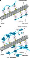
Proposed model for the hyaluronan-microfibril complex mediated by homotypic interaction of VG1Fs. A, Because intact versican binds to HA using its amino-terminal G1 domain, HA is linked to microfibrils through versican molecules by a model identified previously. B, cleavage of the intact versican generates VG1Fs. VG1Fs interact homotypically and with the G1 or G3 domain of versican attached to microfibrils. Consequently, cleaved and recaptured VG1Fs can enhance recruitment of HA to microfibrils.

Similar articles

Cited by

References

    1. Feinberg T., Weiss S. J. (2009) Developmental ECM sculpting. Laying it down and cutting it up. Dev. Cell. 17, 584–586 - PubMed
    1. Kimata K., Oike Y., Tani K., Shinomura T., Yamagata M., Uritani M., Suzuki S. (1986) A large chondroitin sulfate proteoglycan (PG-M) synthesized before chondrogenesis in the limb bud of chick-embryo. J. Biol. Chem. 261, 13517–13525 - PubMed
    1. Zimmermann D. R., Ruoslahti E. (1989) Multiple domains of the large fibroblast proteoglycan, versican. EMBO J. 8, 2975–2981 - PMC - PubMed
    1. LeBaron R. G., Zimmermann D. R., Ruoslahti E. (1992) Hyaluronate binding-properties of versican. J. Biol. Chem. 267, 10003–10010 - PubMed
    1. Zimmermann D. R., Dours-Zimmermann M. T., Schubert M., Bruckner-Tuderman L. (1994) Versican is expressed in the proliferating zone in the epidermis and in association with the elastic network of the dermis. J. Cell Biol. 124, 817–825 - PMC - PubMed

Publication types

MeSH terms

LinkOut - more resources