. 2013 Oct 11;288(41):29414-29.
doi: 10.1074/jbc.M113.472456. Epub 2013 Aug 20.

Dna2 is involved in CA strand resection and nascent lagging strand completion at native yeast telomeres

Affiliations

Dna2 is involved in CA strand resection and nascent lagging strand completion at native yeast telomeres

Martin E Budd et al. J Biol Chem. .

Abstract

Post-replicational telomere end processing involves both extension by telomerase and resection to produce 3'-GT-overhangs that extend beyond the complementary 5'-CA-rich strand. Resection must be carefully controlled to maintain telomere length. At short de novo telomeres generated artificially by HO endonuclease in the G2 phase, we show that dna2-defective strains are impaired in both telomere elongation and sequential 5'-CA resection. At native telomeres in dna2 mutants, GT-overhangs do clearly elongate during late S phase but are shorter than in wild type, suggesting a role for Dna2 in 5'-CA resection but also indicating significant redundancy with other nucleases. Surprisingly, elimination of Mre11 nuclease or Exo1, which are complementary to Dna2 in resection of internal double strand breaks, does not lead to further shortening of GT-overhangs in dna2 mutants. A second step in end processing involves filling in of the CA-strand to maintain appropriate telomere length. We show that Dna2 is required for normal telomeric CA-strand fill-in. Yeast dna2 mutants, like mutants in DNA ligase 1 (cdc9), accumulate low molecular weight, nascent lagging strand DNA replication intermediates at telomeres. Based on this and other results, we propose that FEN1 is not sufficient and that either Dna2 or Exo1 is required to supplement FEN1 in maturing lagging strands at telomeres. Telomeres may be among the subset of genomic locations where Dna2 helicase/nuclease is essential for the two-nuclease pathway of primer processing on lagging strands.

Keywords: DNA Damage Response; DNA Recombination; DNA Repair; DNA Replication; Dna2 Helicase/Nuclease; Exo1 Nuclease; FEN1; MRX; Okazaki Fragment Processing; Yeast.


Figures

FIGURE 1.
FIGURE 1.
5′-CA-strand resection and elongation of a de novo telomere are defective in dna2-1 mutants. A, schematic of experimental design (21). Details are found in the text. B, WT and dna2-1 strains were grown in raffinose-containing media, incubated with nocodazole until 95% of the cells were arrested as dumbbells, collected by centrifugation, and resuspended in medium containing galactose to induce the HO endonuclease and nocodazole to maintain cells in G2 phase. Cells were incubated for 0, 1, 2, 4, and 6 h at 37 °C; chromosomal DNA was isolated, cut with SpeI, electrophoresed on an agarose gel, and blotted to GeneScreen Plus, and the blot was hybridized with the ADE2 probe. The band at about 3 kb is the SpeI fragment spanning the ADE2-HO-LYS2 construct. The ∼0.7-kb fragment corresponds to the SpeI-HO cut fragment. The band at about 1.6 kb represents the endogenous ade2-101 gene and serves as an internal standard to normalize loadings. These experiments were repeated twice with similar results. C and D, dna2-1 cells are deficient in both 3′-GT extension and 5′-CA-strand resection at de novo telomeres. DNA2 and dna2-1 cells were grown, and the HO cut was introduced as in B. Cells were incubated for 0–4 h at 30 or 35 °C, as indicated. Samples were collected at each time point and harvested, and chromosomal DNA was isolated and cut with DdeI, electrophoresed on a 5% denaturing acrylamide gel, electroblotted onto a nylon membrane, and hybridized with an ADE2 riboprobe recognizing sequences adjacent to the 3′-GT-terminated strand (C). The blot was stripped and then rehybridized to a riboprobe recognizing the 5′-CA-terminated strand (D). The DdeI-HO fragment is 300 nt. The 287-nt band below it is the internal standard corresponding to the endogenous ade2-101 locus. C, 3′-GT extension is inhibited in dna2-1. D, 5′-CA-strand resection is defective. Quantification of 300-bp hybridization intensity (Int300) normalized to the 287-bp loading control (Int287) in D is shown in the graph as the mean ± S.E., n = 2. E, resection of the nontelomeric end at the de novo telomere is delayed. UCC5913-2-1 dna2-1 cells were grown in raffinose-containing media at the permissive temperature, 23 °C, incubated with nocodazole until 95% of the cells were arrested as dumbbells, centrifuged, and resuspended in media containing 3% galactose and nocodazole. After incubation for an additional 0–4 h at 30 °C, nonpermissive for dna2-1, cells were collected; chromosomal DNA was isolated, cut with SspI, electrophoresed on a 1% agarose gel, and alkaline-blotted onto GeneScreen Plus. SspI, instead of DdeI used in C, is used to reveal the distal site. Left panel, blot was hybridized with the centromere-distal probe, a PCR product synthesized by oligonucleotides ADH4-HO and NotI-ADH4. The band disappears as resection occurs past the SspI site. Right panel, blot was stripped and reprobed with the proximal probe, a PCR product synthesized using oligonucleotides ADE2–3′ and ADE2–5′. The upper band in the doublet represents the internal ade-102 gene, which should not change, and serves as an internal standard. The two SspI-ADE2 bands (proximal) are more difficult to separate than the two DdeI-ADE2 (proximal) bands that are seen in C.
FIGURE 2.
FIGURE 2.
Analysis of single-stranded GT-overhangs at native telomeres in dna2Δ strains. A, schematic of Y′ telomere. B, determination of overhang extension in S phase. WT (BY1408), pif1Δ (MB121), and dna2Δ pif1Δ (MB161B) were grown at 30 °C, arrested with α factor, and then released into a synchronous cell cycle. Samples were taken at 0, 30, and 45 min after release. DNA was prepared and cleaved with XhoI to release terminal fragments. The fragments were separated by gel electrophoresis and blotted to GeneScreen Plus under neutral/NATIVE conditions as described under “Experimental Procedures.” The blots were probed with a telomere-specific CA-rich oligonucleotide probe, which detects GT-overhangs. All samples were from the same gel but were rearranged in Photoshop for logical presentation. Notice, for instance, seam between samples 5 and 6 and 6 and 7. DENATURED, the same samples (0.1 volume) were analyzed after alkaline blotting as a loading control for XhoI-cut DNA. Only the terminal ∼1.2-kb XhoI fragments (i.e. from Y′ telomeres) are shown. Gels were not run long enough to determine telomere length differences in the respective strains. These experiments were repeated three times with the same result. (Flow cytometry of these strains is shown in Fig. 3, C and D). B, single-stranded DNA is at the telomere termini and is not internal. Analysis of XhoI-cut DNA from synchronized S phase cells was after cleavage with Escherichia coli 3′-exonuclease I. NATIVE, cells were arrested with α factor and released into the cell cycle. S phase samples with GT-overhangs were taken from WT and pif1Δ cells at 35 and 45 min, and samples were taken from dna2Δ pif1Δ at 40 and 55 min to account for the slower progression through the cell cycle of the double mutant. XhoI-digested DNA samples were divided in half, and one-half was treated with bacterial exonuclease 1 as described under “Experimental Procedures.” Samples were then analyzed by neutral blotting as in A (NATIVE). DENATURED, loading control. The same samples (0.1 volume) were analyzed after alkaline blotting. Note that the gel was not run long enough to provide sufficient resolution to estimate telomere length in the mutants, which we have reported previously. C, dna2Δ rad9-320 strains are defective in producing long single-stranded overhangs at native telomeres. The experiment was performed as described in A and B. The NATIVE gel was hybridized to the CA probe. The DENATURED loading control was hybridized to a Y′-specific probe to avoid differences in signal due to different telomere lengths in the various strains. The amount of single-stranded DNA was normalized to the loading control and zero time point. D, quantification of data in C. Note that lanes 10–12 are a repeat of lanes 7 and 8 using a different clone of the same strain and show similar results. E, flow cytometry of samples analyzed in C and D.
FIGURE 3.
FIGURE 3.
dna2 mre11-nuclease-deficient double mutants are not defective in production of single-stranded DNA in S phase at native telomeres. A, top panel, indicated strains were grown at 30 °C, arrested with α factor, and released into the cell cycle as described under “Experimental Procedures.” Samples were collected at the times indicated, cleaved with XhoI, and analyzed by neutral blotting for single-stranded DNA (NATIVE) and alkaline blotting as a loading control (DENATURED) as described in Fig. 2 and under “Experimental Procedures.” Hybridization was carried out with the same probe for both gels. B, indicated strains were treated as in A. C and D, flow cytometric analysis of cell cycle progression of strains used in A and B. E, quantification of overhang data, mean ± S.E., n = 3 for WT, dna2Δ pif1Δ, dna2Δ pif1Δ mre11-H125N; n = 2 for pif1Δ dna2Δ mre11-H56N. The amount of single-stranded DNA in each was normalized to the loading control rehybridized with a Y′-specific probe. Normalization was to 0 time.
FIGURE 4.
FIGURE 4.
Long GT-overhangs appear in S phase on linear YLpFAT10-TEL in dna2, mre11-nuclease minus, and in dna2 mre11-nuclease minus double mutants. A, schematic map of YLpFAT10-Tel, about 7.5 kb plus the length of the telomeric repeats. B, overhangs on YLpFAT10-Tel. The respective strains carrying linear YLpFAT10 were grown at 30 °C, arrested with α factor, and released into the cell cycle for 45 min. Upper panel, genomic DNA was isolated and separated on a neutral gel. In-gel hybridization with the CA probe hybridizing to GT-overhangs was carried out as described (6). The gel was then soaked in NaOH to denature the DNA and, after neutralization, rehybridized with the CA probe to determine the total GT DNA. Lanes 1 and 2, MRE11 DNA2 pif1Δ, 0 and 45 min after release from α factor; lanes 3 and 4, mrell-H125N pif1Δ, 0 and 45 min; lanes 5 and 6, dna2Δ pif1Δ, 0 and 45 min; lanes 7 and 8, mre11-H125N dna2Δ pif1Δ, 0 and 45 min. C, to estimate telomere length and to monitor telomeric DNA on both ends of the plasmid, the same samples were cut with NsiI and separated by gel electrophoresis. Southern blots were hybridized with the CA oligonucleotide (top panel). The blot was then washed and rehybridized with a LEU2 probe to the 2.5-kb end as loading control, copy number control, and length control (middle panel). Finally, the blot was washed and hybridized to the AMP4 probe to confirm the 3.5-kb end (bottom panel). Lanes 1 and 2, pif1Δ, 0 and 45 min after release from α factor; lanes 3 and 4, mrell-H125N pif1Δ, 0 and 45 min; lanes 5 and 6, dna2Δ pif1Δ, 0 and 45 min; lanes 7 and 8, mre11-H125N dna2Δ pif1Δ, 0 and 45 min. These experiments were duplicated (using independent transformants) with the same quantitative results for GT-overhangs, steady-state GT tract length, and steady-state plasmid copy number. D, loading control from chromosomal DNA for C. The blot shown in C was hybridized to a RAD9 probe to normalize for single copy DNA content.
FIGURE 5.
FIGURE 5.
dna2 exo1 mutants accumulate single-stranded DNA in S phase at native telomeres. Analysis of XhoI cut DNA from asynchronous cells. NATIVE, wild type (MB214), cdc13-1 (MB215), exo1Δ (MB216), dna2-2 (MB214), and dna2-2 exo1Δ (MB217) were grown to log phase in medium containing 0.5 m sorbitol, which is essential to allow growth of the dna2-2 exo1Δ mutant and shifted to 37 °C for the indicated times (0, 2, or 4 h) in the absence of sorbitol. DNA was prepared, and GT-overhangs were analyzed on neutral agarose gels as described under “Experimental Procedures” and the legends to Figs. 2 and 3. DENATURED, the same DNA samples were analyzed on a separate gel, which was blotted under denaturing conditions. The experiment shown is a representative example of three biological replicates showing similar results. Quantification of the experiment shown is at the right. The mean of normalization to two different Y′ bands, one at 1.2 kb, is shown, and another at 6.5 kb (data not shown) is found in this strain using the Y′-specific probe.
FIGURE 6.
FIGURE 6.
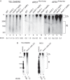
Both dna2-1 and dna2Δ rad9-320 mutants are defective in nascent DNA maturation at telomeres. A, comparison of nascent telomeric DNA in cdc9-1 and dna2-1 mutants. Wild type, cdc9-1, or cdc9-1 rad9Δ mutants were synchronized with α factor and released into S phase for 45 min (WT) or 60 min (cdc9-1 and cdc9-1 rad9Δ) at 36 °C. Genomic DNA was isolated and subjected to alkaline gel electrophoresis to release newly synthesized DNA. Nascent DNA was identified with a telomeric probe (left panel) or an ARS1 probe (right panel), specific for the lagging strand at ARS1, after Southern blotting. The telomere-specific probe was GT-rich, because the nascent DNA from the lagging strand is CA-rich. Ch marks chromosomal sized DNA, and RI indicates DNA shorter than 3 kb. dna2-1 and dna2-1 rad9Δ mutant strains were released from α factor and grown for 60 min at 36 °C before DNA isolation to allow for their slower cell cycle progression. Blots were hybridized to telomere-specific GT probe (left panel) or ARS1 lagging strand probes (middle and right panels). These experiments were repeated at least once with similar results. The RI/Ch ratio, reported below the blots, was determined as described in the text, and the values reported are the average of several experiments (lane 1, n = 1; lane 2, n = 1; lane 3, n = 2; lane 4, n = 2; lane 5, n = 2. B, dna2Δ mutants are defective in nascent DNA maturation at telomeres. The indicated strains were grown at 30 °C after α factor release but were otherwise treated as in A and probed with a telomeric GT-rich probe. Left panel, lanes 1–3, or an ARS1 lagging strand probe; right panel, lanes 4–6. Results are the average of n = 2.
FIGURE 7.
FIGURE 7.
Telomere DNA replication and the proteins involved. A replication fork is initiated in the subtelomeric region creating forks that replicate templates (black lines) outward toward the telomere. The GT-rich strand uses the leading strand polymerase, and the CA-strand uses the lagging strand polymerase. Left leading strand, synthesis by the leading strand polymerase results in a chromosome with a blunt end. This end is analogous to the HO endonuclease cut chromosome in the de novo telomere assay used in this study. The appearance of the normal 3′-GT-overhang on the leading strand therefore requires 5′- to 3′-nuclease processing. Subsequent binding of Cdc13 would allow telomerase binding and synthesis. Nucleases MRX, Sae2, Dna2, and/or Exo1 may be required for efficient 5′- to 3′-CA resection (, and this work). Right lagging strand, pol δ (lagging strand polymerase) arrives at the telomere significantly later than pol ϵ (leading strand polymerase) (90). After leading strand replication is completed, the helicase presumably falls off, and the leading and lagging strand syntheses are uncoupled. This supports the idea that a 3′-GT-overhang, having at least the size of an Okazaki fragment, appears on the chromosome synthesized by the lagging strand. Further evidence suggesting that a 3′-GT-overhang arises on the lagging strand during its replication is that in an in vitro SV40 linear DNA replication assay, leading strand synthesis replicates the DNA to the end but lagging strand synthesis leaves a 500-bp single-stranded gap at the end (102). The appearance of RPA at telomeres coincides with the arrival of pol ϵ suggesting that RPA binds the GT template. In mammalian cells, a special process replaces RPA with telomere-specific DNA-binding proteins that do not activate the checkpoint, and we propose the same occurs in yeast for RPA and Cdc13 (103, 104). These overhangs recruit telomerase, recruit protective telomere capping proteins, such as Cdc13, and themselves can form protective structures such as t-loops (13, 105, 106). 3′-GT tails are the primer for synthesis by telomerase.

Similar articles

Cited by

References

    1. Shampay J., Szostak J. W., Blackburn E. H. (1984) DNA sequences of telomeres retained in yeast. Nature 310, 154–157 - PubMed
    1. Walmsley R. W., Chan C. S., Tye B. K., Petes T. D. (1984) Unusual DNA sequences associated with the ends of yeast chromosomes. Nature 310, 157–160 - PubMed
    1. Larrivée M., LeBel C., Wellinger R. J. (2004) The generation of proper constitutive G-tails on yeast telomeres is dependent on the MRX complex. Genes Dev. 18, 1391–1396 - PMC - PubMed
    1. Wellinger R. J., Wolf A. J., Zakian V. A. (1993) Saccharomyces telomeres acquire single-strand TG1–3 tails late in S phase. Cell 72, 51–60 - PubMed
    1. Wellinger R. J., Zakian V. A. (2012) Everything you wanted to know about Saccharomyces cerevisiae telomeres: beginning to end. Genetics 191, 1073–1105 - PMC - PubMed

Publication types

MeSH terms